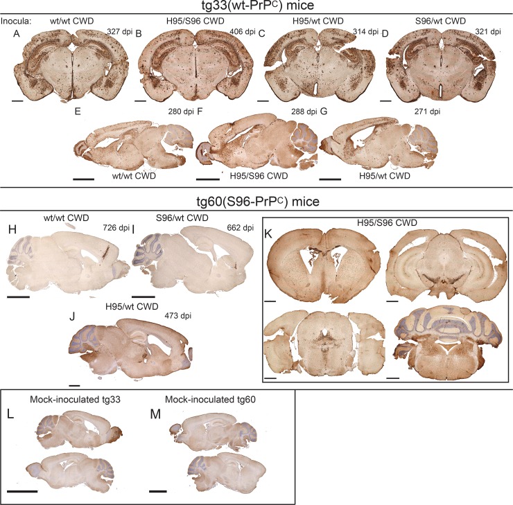
FIG 3.
Distribution of PrPCWD aggregates in the brains of tg-deer-PRNP mice inoculated with different white-tailed deer CWD allotypes. (A to G) PrPCWD aggregates in the brains of tg33 mice inoculated with different white-tailed deer CWD allotypes. (H and I) Abnormal PrP aggregates were detected after >700 dpi in the brains of tg60 mice without clinical signs inoculated with the wt/wt or S96/wt CWD allotypes. (J and K) Only tg60 mice inoculated with H95+ CWD allotypes had clinical signs and were consistently positive for PrPCWD aggregates. (K) Coronal brain sections from a clinically ill tg60 mouse infected with H95+ CWD prions (414 dpi). (L and M) Brain sections of mock-infected tg33 and tg60 mice. Bars, 1 mm (A to D, J, and K), 2.5 mm (E to I and M), and 5 mm (L). Tissue sections were stained with anti-PrP monoclonal antibody BAR224.

