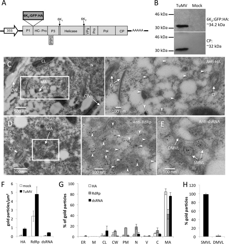
FIG 4.
Subcellular localization of TuMV RNA replication sites. (A) Schematic representation of the infectious clone TuMV/6K2:GFP:HA that coexpresses 6K2 as a GFP:HA protein fusion. Black lines, the plasmid backbone; arrow, the cauliflower mosaic virus 35S promoter; AAAAA, the position of the polyadenylated tail; rectangles, TuMV proteins. 6K2:GFP:HA is inserted between P1 and HCpro. PIPO, pretty interesting Potyviridae ORF. (B) Western blot analysis of 6K2:GFP:HA and CP expression in N. benthamiana plants after TuMV/6K2:GFP:HA infection. (C to E) Immunogold labeling was performed on the cross sections of mock- and TuMV-infected N. benthamiana leaf tissues by using anti-HA, anti-RdRp, and anti-dsRNA antibodies. Higher-magnification images of the areas in the white rectangles are shown on the right of each panel. Arrowheads, HA-specific (C) and RdRp-specific (D) gold particles, which are located in TuMV-induced membranous aggregates. (E) The dsRNA-specific gold particles are mainly localized to TuMV-induced SMVL structures. (F to H) The number of gold particles per square micrometer in mock-infected versus TuMV-infected cells (F), the relative labeling distribution in infected cells (G), and the relative labeling distribution in TuMV-induced membranous aggregates (H) are shown. The results of two different labeling experiments were considered, and 200 gold particles were counted for each experiment. N, nucleus; CL, chloroplast; CW, cell wall; MA, membranous aggregate; CI, cytoplasmic inclusion body; SMVL, single-membrane vesicle-like structure; DMVL, double-membrane vesicle-like structure; ER, endoplasmic reticulum; M, mitochondrion; PM, plasma membrane; C, cytosol; V, vacuole.

