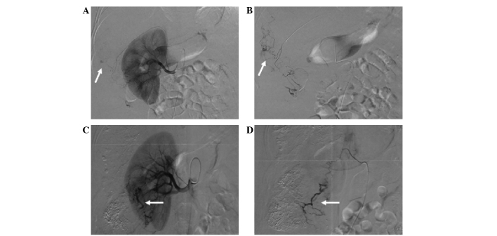
Figure 11.
Hepatocellular carcinoma supplied by the renal artery. (A) Angiogram of a prominent superior renal capsular artery shows a hypervascular tumor (white arrow). (B) Renal arteriogram shows the hepatic tumor (white arrow) with a microcatheter inserted into the branch of the renal artery. (C) Right renal arteriogram shows staining of the tumor (white arrow) supplied by the renal artery. (D) A further selective angiogram shows tumor staining (white arrow).

