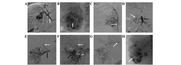
Figure 7.
Development of extrahepatic arteries after several sessions of transcatheter arterial chemoembolization (TACE). (A) After the third session, the hepatic artery was occluded. The right inferior phrenic artery (arrow) supplied the tumor. (B) Embolization with iodized oil (arrow) through the right inferior phrenic artery. (C) The right inferior phrenic artery (arrow) supplied the tumor. (D) Hepatocellular carcinoma (HCC) supplied by a branch (white arrow) of the superior mesenteric artery (black arrow) after the fourth session of TACE. (E) The right adrenal artery (white arrow) supplied the partial tumor after the sixth session of TACE (black arrow indicates the renal artery). (F) HCC supplied by a branch (white arrow) from the pancreaticoduodenal arterial arch (black arrow; black arrow head indicates the superior mesenteric artery). (G) After the sixth session of TACE, the HCC was supplied by the left gastric artery (black arrow). (H) Embolization with iodized oil (white arrow) through the left gastric artery.

