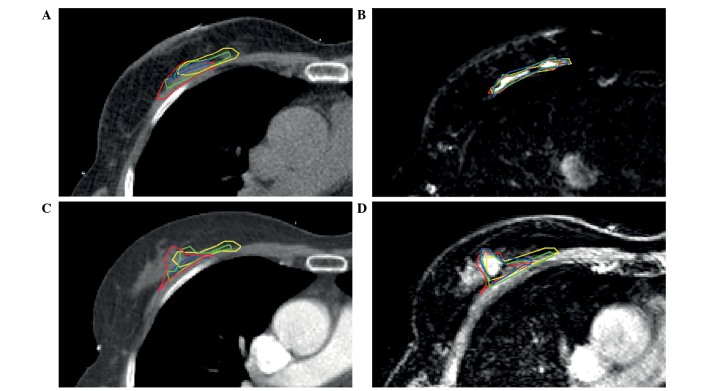
Figure 1.
CT and MRI scans of a 64-year-old patient with pT1cN0(sn) ductal carcinoma of the right breast. The cavity visualization score was 2 on CT. CT-guided tumor bed delineation was determined by four observers, demonstrated in different colors, on the same transverse slice on (A) postoperative CT, (B) postoperative T2-weighted MRI, (C) preoperative CE-CT and (D) preoperative CE-MRI scans. CT, computed tomography; MRI, magnetic resonance imaging; CE, contrast enhanced.

