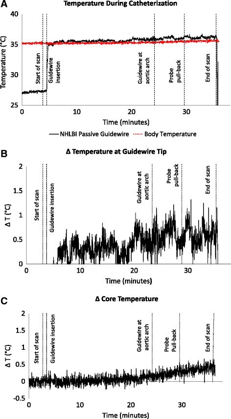
Fig. 7.

Temperature during left heart catheterization in swine. The panels depict three simultaneous tracings during bSSFP with a flip angle of 45°. First scanning begins, then the guidewire is advanced retrograde to the aortic arch, and then a temperature probe is withdrawn alongside the guidewire in order to measure tip and shaft temperature. a shows the guidewire temperature probe (black) and simultaneous core body temperature (red). On a narrower scale, (b) shows the instantaneous difference between the guidewire and core body temperature, while (c) shows the core body temperature rise during CMR. The guidewire temperature rise was negligible
