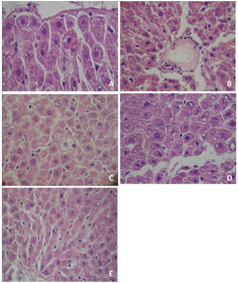
Figure 1.
Histological analysis of rat liver tissues. Tissues were stained with hematoxylin-eosin and analyzed using a light microscope and a 400× oil immersion objective. (A) control group; (B) Pentylenetetrazol administration produced infiltration of inflammatory cells (IIC); (C) Pentylenetetrazol administration produced hepatic steatosis (HS); (D) organic yerba mate treatment (50 mg/kg) plus pentylenetetrazol prevented infiltration of inflammatory cells (IIC) and hepatic steatosis (HS); (E) conventional yerba mate treatment (50 mg/kg) plus pentylenetetrazol prevented IIC and HS. (A, D and E: normal hepatocytes). The photomicrographs show the most representative slide of each group.

