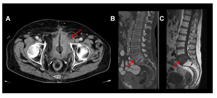
Figure 1.
Large bladder tumor visualized by contrast enhanced computed tomography (CT) reaching the acetabulum on the left side and infiltrating the perivesical fatty tissue (A). Note there is an additional spinal metastasis in L5 (B, sagittal reformatted CT), seen as a destructive lesion in L5 with sintering of the vertebra. This is confirmed by MRI: the lesion is seen hypointense in T1w, confirming infiltration of the bone marrow by tumor tissue (C, T1w sagittal).

