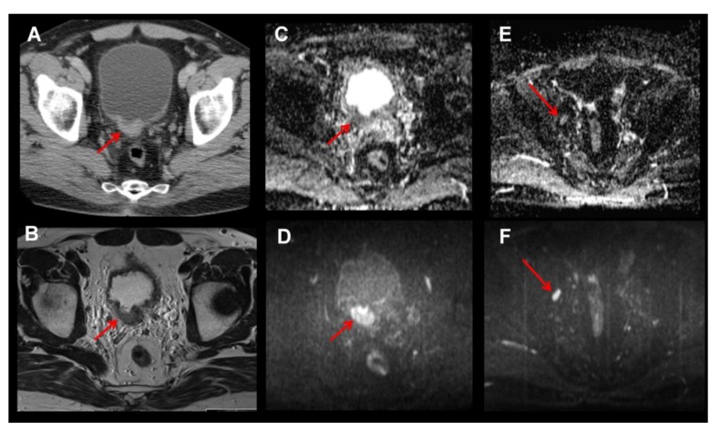
Figure 2.
Bladder tumor at dorsal bladder wall visualized by CT as a wall thickening with contrast enhancement (A). Of note, soft tissue contrast is superior in MRI compared to CT (B, T2w axial). Additional diffusion weighted imaging (DWI; C–F) improves delineation of the tumor mass as a hyperintense mass in the b-value images (D, b800 image) and allows evaluating apparent diffusion coefficient (ADC) values (C) as potential marker of tumor cellularity. Moreover, DWI might be helpful for evaluating lymph node metastasis, as metastatic lymph nodes usually also show lower ADC values (F, b800 image showing a hyperintense iliacal lymph node on the right; E, low ADC value suggests malignancy).

