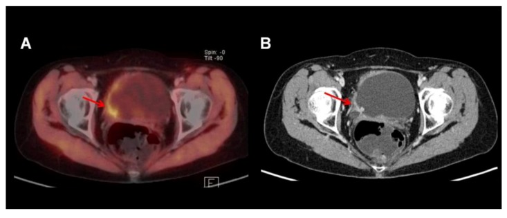
Figure 3.
Bladder tumor with tracer uptake in the right posterior-lateral bladder wall as visualized by 11Choline-PET/CT. Note that there is excellent delineation of tumor and bladder lumen due to the fact that 11C-Choline is usually not excreted by the urine (A: fused dataset). However, anatomical detail is superior in the contrast enhanced CT part of the PET/CT (B). Note the hypervascularized tumor involving the right ureteral orifice with consecutive dilatation of the right ureter.

