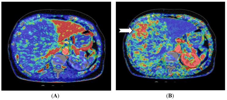
Figure 3.
CT perfusion examination of a 77-year-old female after right-sided portal vein embolization prior to liver resection. The patient has a large HCC in the right liver lobe and segment 4. (A) Perfusion shows the portal flow, which is eliminated on the right side and elevated in the left liver lobe; (B) Perfusion index (Arterial Flow/Arterial Flow + Portal Flow). This index is low in the left side due to elevated portal flow, and the index is high in all of the embolized segments, but highest in the vascular part of the HCC (arrow) (Images reconstructed with Vitrea 6.2, Vital Images A Toshiba Medical Systems Group).

