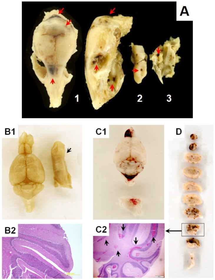
Figure 2.
Development of hemorrhagic vasculopathy in irradiated B6D2F1/J mice at the mortality period. Gross pathology and histopathology assessment (hematoxylin and eosin staining, i.e., H & E) of intracranial hemorrhage in moribund B6D2F1/J mice subjected to hARS. Panel A. (1) Images a mouse skull: dorsal plane (left) and lateral plan (right); (2) Image of tongue; (3) Image of a fragment of skull shown in (1). Brownish areas of extravasated blood are indicated with red arrows. Panels B1 and B2 are specimens from a sham animal. Tongue is indicated with a black arrow in B1. Panels C1, C2, and D are specimens from an irradiated animal. A fragment of cranium is indicated with a black arrow in C1 where the presence of epidural hemorrhage is observable. Panels B2 and C2 display H & E-staining images of coronal sections through the cerebellar cortex. Panel D displays gross-image of coronal sections through entire brain. As shown in C1 and D subdural and interstitial hemorrhage randomly occurred in different part of the brain; predominantly affecting cerebellum and olfactory. Experimental conditions: as indicated in the Figure 1.

