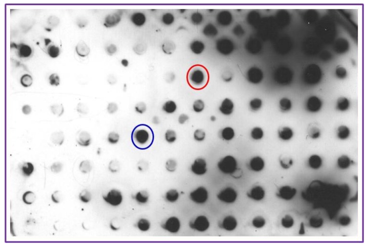
Figure 2.
Hybridoma screening for capture and detection antibody selection: LPGDS (pure protein 20 ng) was transferred to PVDF membrane using a Bio-Rad dot blot apparatus and probed with ascitic fluid generated from culturing different hybridoma clones against LPGDS. The membrane was developed using ECL detection kit. Dark dots indicate positive interaction. Positive antibody from red circle was designated as Antibody 1 (AB1) and that in the blue circle is designated as Antibody 2 (AB2) for use in the biomarker detection method. Other clones showed specificity at different levels and these can also be used to detect LPGDS. AB1 and AB2 were selected based on their strong interaction with LPGDS.

