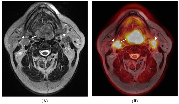
Figure 3.
A 65-year old male patient with lingual carcinoma (A) T2-weighted MRI and (B) 18F-FDG PET/MRI. The primary tumour (arrows) and nodal metastases (arrowhead) are considerably more conspicuous on PET/MRI compared with T2-weighted images. This advantage could be exploited in detecting small occult head and neck tumours.

