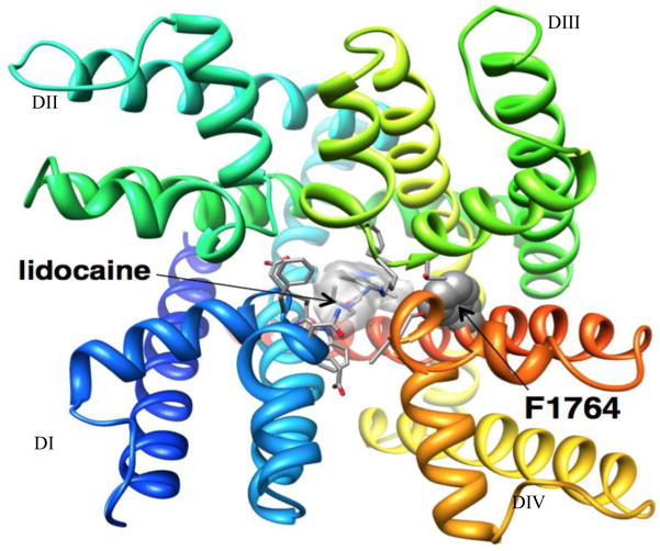
Figure 1.
Docking of lidocaine in the Rosetta model of Nav1.5 channel pore. View of Nav1.5 - lidocaine model from the extracellular side of the membrane. Each domain is colored individually and labeled. Side chains of key residues for lidocaine binding are shown in space-filling and stick representation (Yarov-Yarovoy Laboratory).

