
Figure 3.
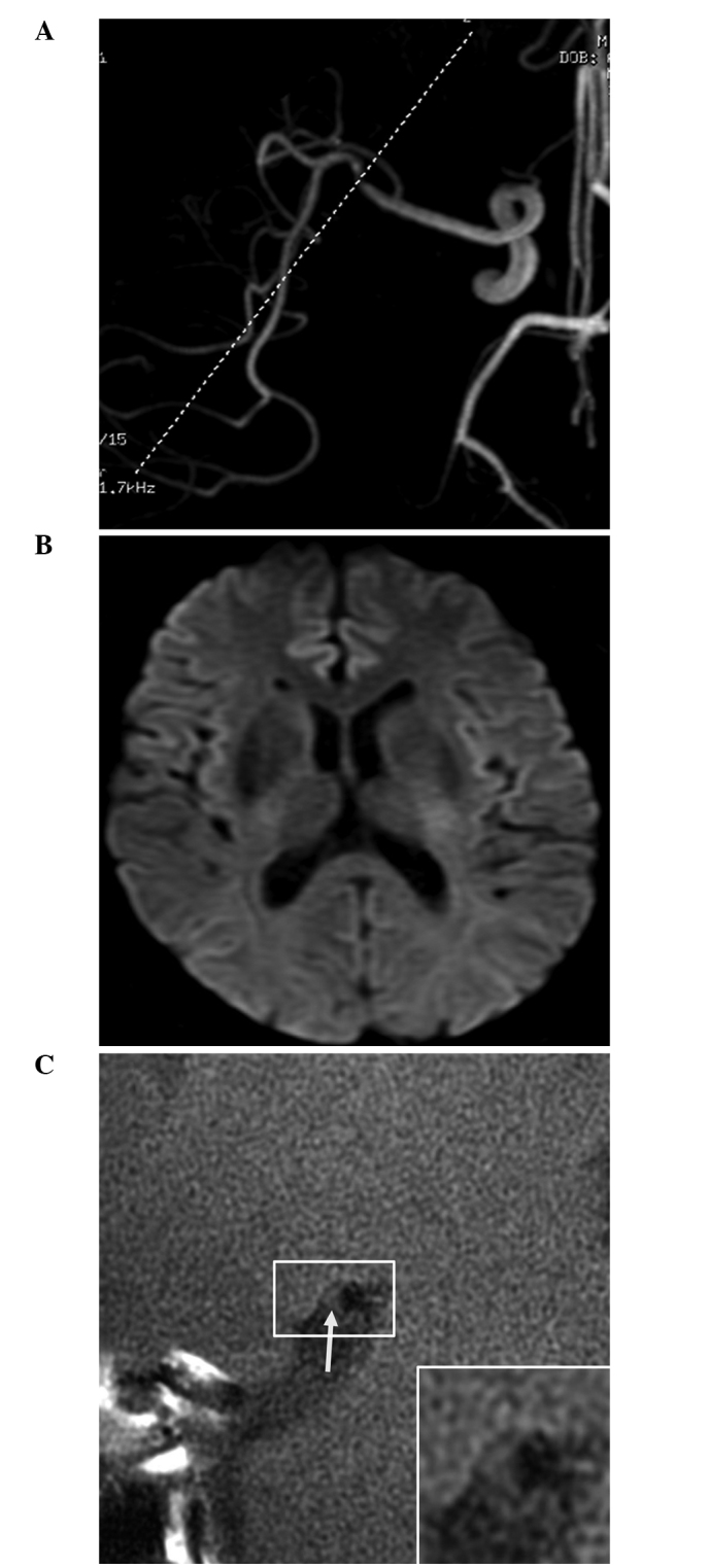
Magnetic resonance angiography (MRA) and high-resolution magnetic resonance imaging (HR-MRI) results of a 64-year-old male patient with non-acute cerebral infarction. (A) MRA revealed marked stenosis of the distal M1 segment of the right middle cerebral artery; (B) Diffusion-weighted imaging revealed no abnormal high signal; and (C) HR-MRI revealed no plaque enhancement (arrow). The remodeling rate of the stenotic lumen was 0.89.