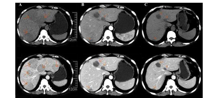
Figure 1.
Differences observed in the upper abdominal CT scan images performed prior and subsequent to lenalidomide therapy. (A) non-enhanced (top) and enhanced (bottom) CT preceding lenalidomide therapy, performed February 25th 2014. Multiple round-shaped and well-defined lesions of different sizes with low attenuations were present in the hepatic parenchyma on non-enhanced CT. Following the administration of contrast reagents, the enhancements of the lesions were mild and heterogeneous. (B) Non-enhanced (top) and enhanced (bottom) CT subsequent to 4 cycles of RCD regimen therapy (July 1st 2014). A reduction in the number and size of the round-shaped lesions in the hepatic parenchyma was observed, compared with prior to treatment. (C) Non-enhanced (top) and enhanced (bottom) CT following 5 cycles of RCD regimen (August 25th 2014). Further reduction in the size of the round-shaped lesions present in the hepatic parenchyma was observed, although the lesions did not disappear completely. CT, computed tomography; RCD regimen, lenalidomide, 25 mg, days 1–21; cyclophosphamide, 50 mg, days 1–21; and dexamethasone, 20 mg, days 1, 8, 15 and 22; 28 days/cycle.

