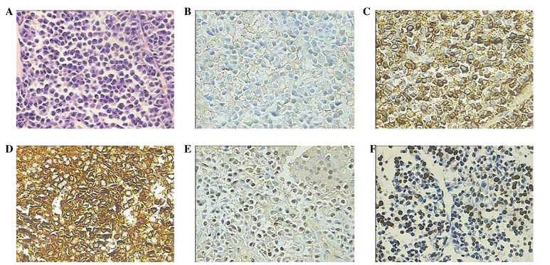
Figure 3.
Histopathological features of the percutaneous needle biopsy of the hepatic lesion. (A) Hematoxylin and eosin staining identified the presence of plasma cells. (B) Immunohistochemical staining demonstrated that the plasma cells were negative for CD20, but positive for myeloma markers (C) CD38 and (D) CD138 and partially positive for (E) multiple myeloma oncogene 1. (F) In situ hybridization for Epstein-Barr virus-encoded RNA was positive. Magnification, ×400. CD, cluster of differentiation.

