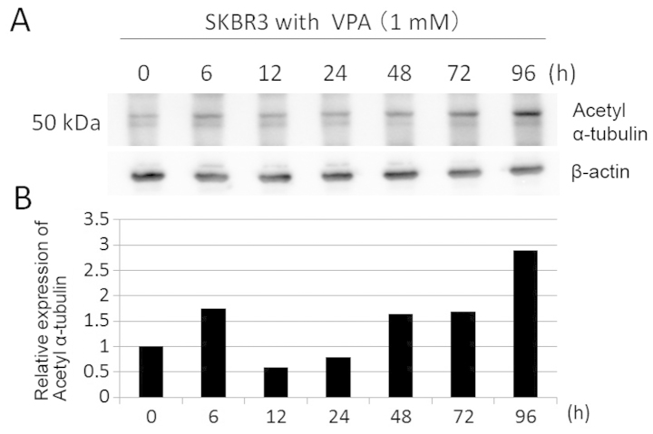
Figure 9.
The effects of VPA on α-tubulin acetylation in SKBR3 cells. (A) The expression levels of acetyl-α-tubulin were detected by immunoblotting protein extracts prepared from SKBR3 cells treated with 1 mM VPA for ≤96 h. The amount of β-actin in each sample was used as the loading control. (B) The expression levels of acetyl α-tubulin were reported in the histogram after normalization against β-actin, relative to the value obtained in the absence of VPA (ratio).

