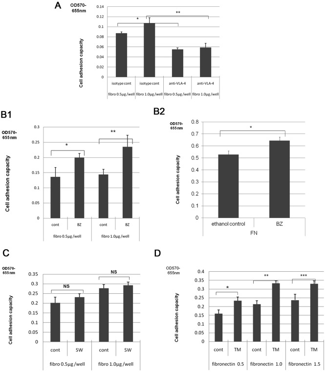
Figure 3.
Effect of alteration of cell surface glycosylation on cell adhesion to fibronectin. The adhesion of cells to fibronectin, and the effect of anti-VLA-4 antibodies or alteration of cell surface glycosylation on this adhesion were assayed using fibronectin coated culture plates. Adhesion was monitored by measurement of absorption at 570–655 nm. (A) Effect of anti-VLA-4 or isotype control (cont) antibodies on HBL-8 3G3 cloned cell adhesion to plates coated with the indicated concentrations of fibronectin (fibro) (*p=0.0003, **p=0.0035). (B) Effect of BZ or control (cont) treatment on adhesion to fibronectin (fibro) of HBL-8 3G3 cells (B-1) (*p=0.0327, **p=0.0194) or of H-ALCL cells (B-2) (*p=0.004). Data shown are representative of two independent experiments performed in triplicate. (C) Effect of SW or control (cont) treatment on adhesion to fibronectin (fibro) of HBL-8 3G3 cells (NS, not significant). Data shown are representative of three independent experiments performed in triplicate. (D) Effect of TM or control (cont) on HBL-8 3G3 cloned cell adhesion to fibronectin (*p=0.0136, **p=0.0010, ***p=0.0126). Data shown are representative of two independent experiments performed in triplicate. p-values were calculated based on Student's t-test.

