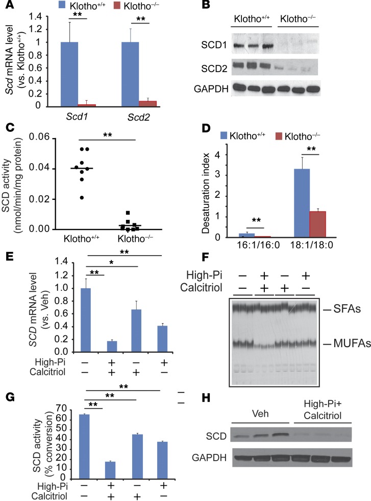
Figure 1. Klotho deficiency reduces the expression of SCD1 and SCD2 in VSMCs.
Eight-week-old male SMMHC-GFP; Klotho–/– (Klotho–/–) mice and SMMHC-GFP; Klotho+/+ (Klotho+/+) mice (n = 8) were sacrificed after a 4-hour fasting. (A) Levels of Scd1 and Scd2 mRNA in the aortas of SMMHC-GFP; Klotho–/– mice and SMMHC-GFP; Klotho+/+ mice. VSMCs were isolated as GFP+ cells by immunomagnetic cell sorting. Scd1 and Scd2 RNA expression was determined by qPCR. (B) Immunoblot analysis of SCD1 and SCD2 protein. Total protein extract was prepared from the aortas and subjected to immunoblot analysis with SCD1- and SCD2-sepcific antibodies. (C) Microsomal SCD activity in the aortas. Microsomal protein (100 μg) was incubated with 14C-18:0-CoA in the presence of NADH. SCD activity was determined as the conversion of 18:0-CoA to 18:1n-9. (D) Desaturation index in the medial layer of aortas from SMMHC-GFP; Klotho–/– mice and SMMHC-GFP; Klotho+/+ mice. Levels of fatty acids were determined by gas chromatography analysis. (E) High-phosphate and calcitriol reduced SCD mRNA in human VSMCs. (F) Autoradiography and (G) quantification of SCD activity in VSMCs treated with high-phosphate (Pi) and calcitriol. (H) Combination of high-phosphate and calcitriol reduced levels of SCD protein. Human VSMCs were treated with 2.0 mM phosphate and 100 nM calcitriol for 7 days. **P < 0.001 vs. Klotho+/+ mice (2-tailed Student’s t test).

