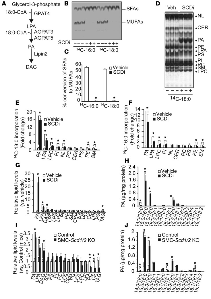
Figure 5. SFAs were preferentially incorporated into PA and accumulated as fully saturated PAs in VSMCs in vitro and in vivo.
(A) Schematic representation of GPAT4, AGPAT3, and AGPAT5, which are localized in the ER. (B) Autoradiography and (C) quantification of SCD activity in human VSMCs treated with 300 nM of the SCDi CAY10566 using 14C-18:0 and 14C-16:0 as substrates (n = 6). VSMCs were treated with 14C-18:0 and 14C-16:0 for 6 hours in the presence/absence of SCDi. SFAs and MUFAs isolated from total cell lysate were separated on a silver nitrate–coated TLC. (D) Autoradiography and quantification of (E) 14C-18:0 and (F) 16:0 incorporation into the lipid fraction in human VSMCs treated with SCDi. Human VSMCs were pretreated with SCDi for 2 hours and incubated with 14C-18:0 and 14C-16:0 for 6 hours in the presence/absence of SCDi. Total lipids isolated from total cell lysate (3 mg protein) were separated on a boric acid–coated TLC. (G) LC-MS–based lipidomic analysis and (H) absolute levels of PA species in VSMCs treated with SCDi. Human VSMCs were treated with SCDi for 12 hours. Lipid content was quantified with LC-MS/MS. (I) LC-MS–based lipidomic analysis and (J) absolute levels of PA species in the aortic medial layers of SMC-Scd1/2 KO mice. Mice (n = 6) were sacrificed at 18 weeks old. The medial layer of aortas were dissected under a dissecting microscope. LPE, lysophosphatidylethanolamine; NL, neutral lipids. *P < 0.01 (2-tailed Student’s t test).

