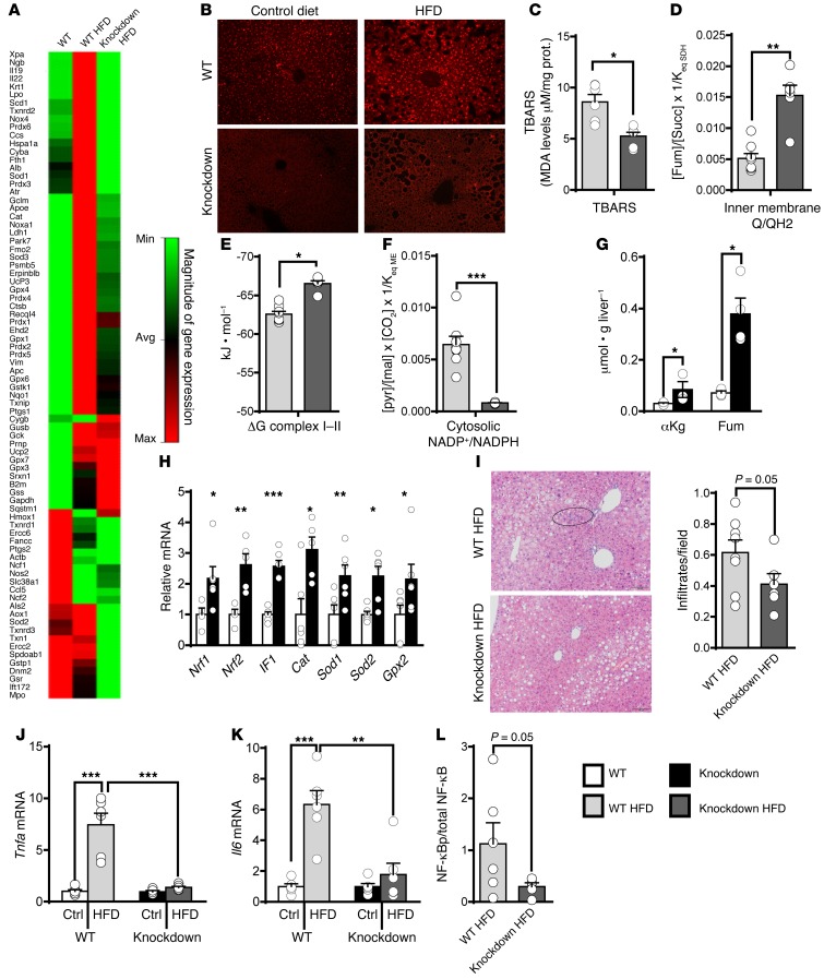
Figure 6. Preventing the induction of anaplerosis/cataplerosis protected against hepatic oxidative stress and inflammation during a HFD.
Oxidative stress, indicated by (A) a qPCR gene array, (B) histological DHE ROS staining, and (C) lipid peroxidation (n = 6), was increased by a HFD in WT, but not knockdown mice. TBARS, thiobarbituric acid reactive substances. (D) The Q/QH2 ratio, estimated from the fumarate/succinate ratio, was oxidized in knockdown mice (n = 6–8). (E) The calculated free energy of complexes I and II was more negative in knockdown mice (n = 6–8). (F) The NADP+/NADPH ratio, estimated from the pyruvate/malate ratio, was reduced in knockdown mice (n = 6–8). (G) TCA cycle intermediates with antioxidant properties were increased in knockdown mice (n = 3–4) as were (H) antioxidant genes (n = 6). Knockdown mice were protected from the inflammatory response of a HFD as indicated by (I) fewer inflammatory infiltrates in H&E-stained tissue (n = 6–8) and lower expression of (J) Tnfa and (K) Il6 mRNA (n = 6). (L) NF-κB S536 phosphorylation was reduced in knockdown liver (n = 5–6). Original magnification, ×10 (I); ×20 (B). Data are shown as mean ± SEM. Statistical differences were detected by 2-way ANOVA (J and K), 2-tailed t test (C–H), or 1-tailed t test (I and L). *P < 0.05; **P < 0.01; ***P < 0.001.

