Figure 3.
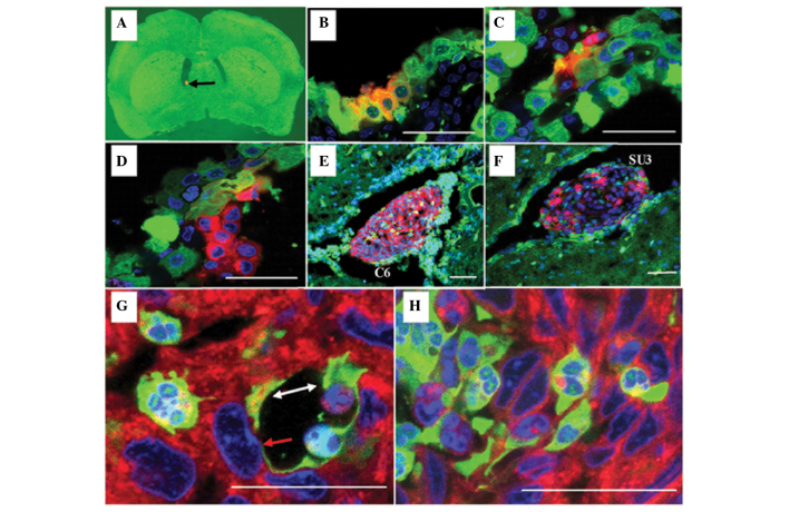
Choroid plexus papillary epithelioma was analyzed using laser confocal microscopy (scale 20 µm). Red fluorescent C6 or SU3 cells were injected into the lateral ventricle of a green fluorescent nude mouse with the assistance of stereotaxic apparatus: (A) Red fluorescent protein (RFP)-expressing tumor cells (black arrow; magnification, 12.5×) settled at the lateral ventricle 1 day following implantation. (B and C) Cell fusions consisting of choroid corpora mammillaria cells and tumor cells 3 days following implantation; some of the choroid corpora mammillaria infrastructure remained (B) normal, whereas other parts were (C) disordered. (D) The tumor predominantly consisted of RFP-expressing tumor cells that projected into the cerebroventricular region 11 days following implantation. (E and F) At the advanced stage of the tumor 15 days following implantation, the tumor appeared large and was accompanied by hydrocephalus. (G and H) Although a few green fluorescent protein-expressing host cells were found in the tumor, the majority were fused polykaryocytes. In particular, mosaic blood vessels were formed by the tumor cells (white arrow; G), and fused host endothelial cells (red arrow; G) could be observed in the choroid plexus.

