Figure 5.
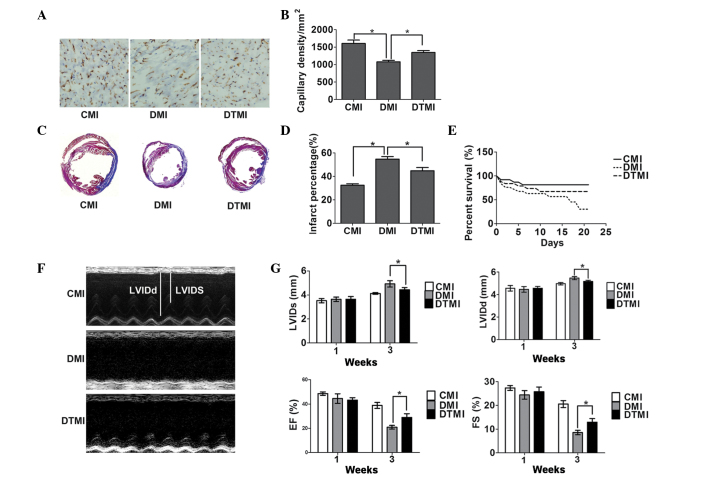
Treatment with the antioxidant Tempol ameliorates cardiac status in type 1 diabetic mice following myocardial infarction (MI). Capillary density, percentage myocardial infarct area and cardiac function were measured using immunohistochemistry, Masson's trichrome staining and echocardiography, respectively, on day 21 after MI. (A) Immunostaining for CD31 (brown) and the cell nucleus (blue) in the border zone of the infarcted myocardium. Scale bar, 100 µm. The number of vessels was measured using Image Pro-Plus. (B) Quantitative analysis of capillary density (n=5). (C) Masson's trichrome staining of the mouse hearts. Scale bar, 3 mm. (D) Percentage infarct area (n=5). (E) Survival rate (n=20). (F) Representative M-mode images. (G) Quantitative analysis of left ventricular internal dimension-systole (LVIDs), left ventricular internal dimension-diastole (LVIDd), fractional shortening (FS) and ejection fraction (EF) (n=5). Data are presented as the mean ± standard error of the mean. *P≤0.05. CMI, control MI group; DMI, diabetic mice with MI; DTMI, DMI group treated with Tempol.

