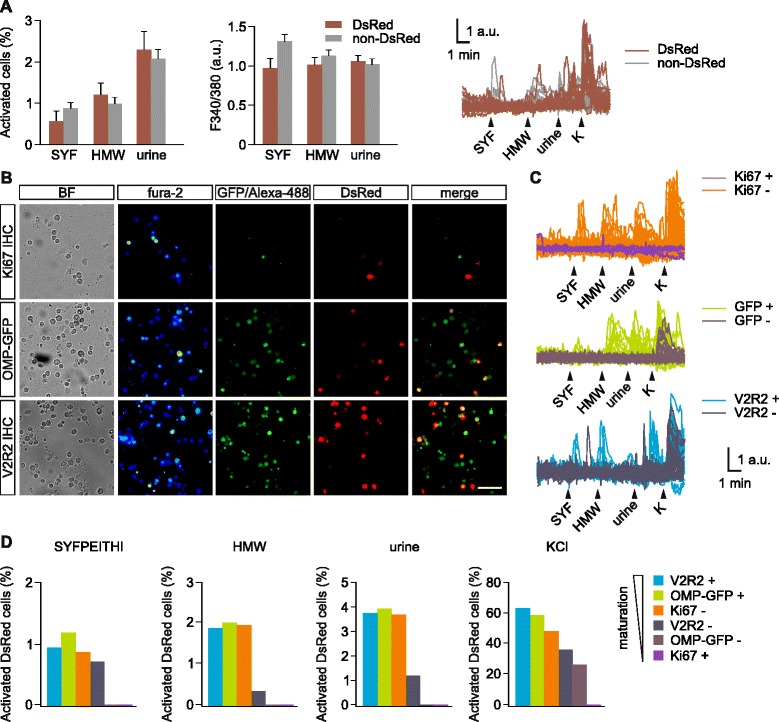
Fig. 3.

Vomeronasal sensory neurons (VSN) activation by pheromonal compounds at different maturity stages. (a) Dissociated vomeronasal organ (VNO) cells from doublecortin (Dcx)-DsRed mice, either DsRed+ (red bars) and DsRed– (gray bars), show intracellular calcium transients in response to the major histocompatibility complex-derived peptide SYFPEITHI (SYF, 10−11 M), whole male urine (1:300 dilution) and its high molecular weight fraction (1:300). Both the number of activated cells (two-way ANOVA: F1,149 = 0.04; P = 0.85) and the intracellular fura-2 fluorescent increases (two-way ANOVA: F1,186 = 1.3; P = 0.26) indicate no differences in the response rate of DsRed positive vs. negative cell populations for all three stimuli. Example traces of Ca2+ transients in single cells in response to the stimuli. High KCl (90 mM) solution was delivered at the end of the experiment to assess cell viability. (b) Post-hoc immunolabeling for Ki-67, V2R2 or endogenous OMP-GFP in Ca2+-imaged VNO cells. Representative images of VNO cells taken with transmission light, bright field, fura-2 pseudocolor fluorescence 340/380 nm ratio (fura-2), 488 nm excitation light of green-labeled secondary antibodies, or OMP-GFP fluorescence (GFP/Alexa-488), DsRed fluorescence (DsRed), and merged ex 488 nm/DsRed color images (merged). OMP-GFP/Dcx-DsRed double transgenic mice were used to label OMP-GFP cells. Scale bar, 50 μm. (c) Representative fura-2 ratio traces of DsRed+ cells labeled with Ki-67, OMP-GFP, or V2R2. Responses of cells positive vs. negative for Ki-67, OMP-GFP, and V2R2 are shown. (d) Percentage of activated DsRed cells classified by marker expression. Near-maximum response rates to the three stimuli are reached in cells positive for V2R2 and OMP and negative for Ki-67. No responses were found in cells positive for Ki-67. Cells negative for OMP-GFP showed responses only to high K+
