
Fig. 4.
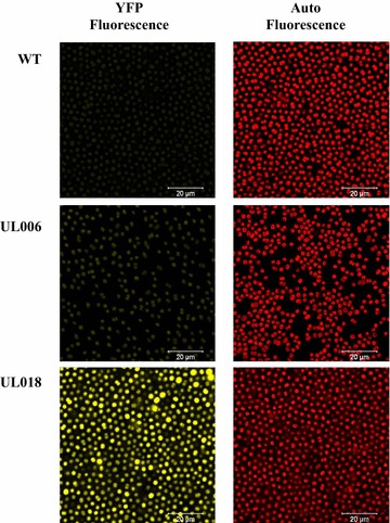
Confocal microscopy analysis of Synechocystis PCC 6803 WT, UL006 and UL018 cells. On the left hand side, representative images of cells enhanced for imaging of YFP, on the right hand side, the same representative images of cells enhanced for imaging of auto-fluorescence of Synechocystis PCC 6803