
Fig. 3.
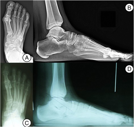
Foot AP and lateral radiographs with standing (a, b Maasai, c, d Korean). The TNCA and T1MTA were significantly greater in the Maasai group on the AP images of the weight-bearing foot, indicating more forefoot abduction related to hindfoot eversion. HVA and IMA were significantly greater in the Korean group. On the lateral images of the weight-bearing foot, the Meary angle and NCO were significantly increased in the Maasai group