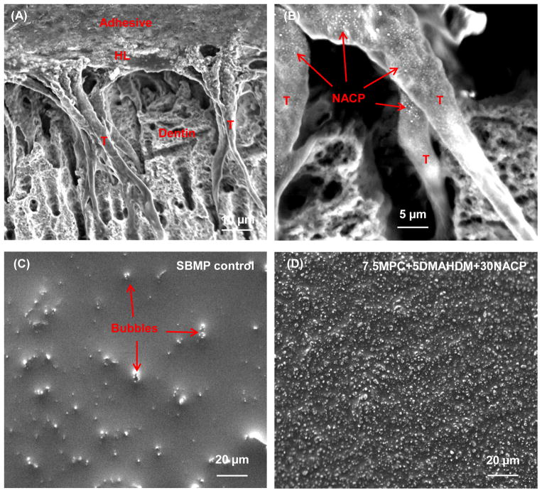
Fig. 3.
Representative SEM images of dentine-adhesive interfaces at the cross-sections, as well as adhesive coating surface textures (without cross-section). (A, B) Cross-section for 7.5MPC+5 DMAHDM+30 NACP group at a lower and higher magnification. The adhesive filled the dentinal tubules and formed resin tags “T”. “HL” indicates the hybrid layer between the adhesive and the underlying mineralized dentine. Arrows in (B) indicate NACP in the dentinal tubules. (C) Surface of SBMP control coating on root dentine, and (D) surface of 7.5MPC+5DMAHDM+30NACP coating on root dentine. In (C), arrows indicate air bubbles in control adhesive coating. In (D), the 7.5MPC+5DMAHDM+30NACP adhesive coating had a solid and dense appearance without air bubbles. The adhesive had completely sealed the root dentine surface.

