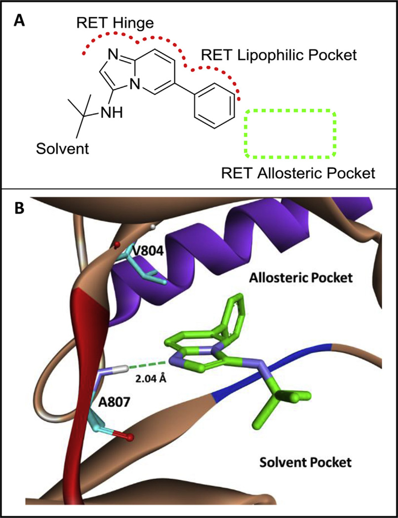
Fig. 1.
A) A novel RET hit (1, RET IC50 ~2.0 mM) from an MCR-based library. The scaffold is predicted to bind the kinase in an atypical, backward binding mode to that of ATP. B) Computational binding of 1 in the RET kinase. Compound 1 is predicted to bind the hinge of RET by hydrogen bonding to A807. The phenyl ring system of 1 accesses a lipophilic pocket close to the RET allosteric pocket. The RET hinge is shown in red, the DFG loop is shown in blue, and the c-Helix is shown in purple. Computational experiments were completed with AutoDock Vina [39]. (For interpretation of the references to colour in this figure legend, the reader is referred to the web version of this article.)

