FIGURE 1:
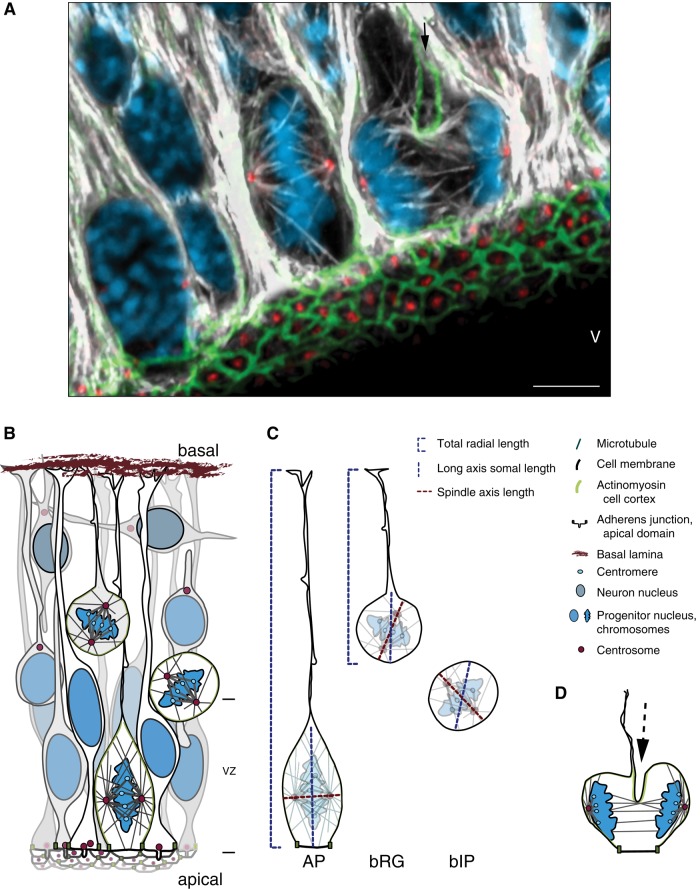
(A) Apical progenitors (APs) dividing at the ventricular surface of the developing cortical wall. Maximum intensity projection of three 0.75-μm optical sections of E14.5 wild-type mouse dorsolateral telencephalon, showing mitotic microtubules in dividing APs. Double immunofluorescence for α- and γ-tubulin combined with 4′,6-diamidino-2-phenylindole (DAPI) and phalloidin staining; DNA in blue, microtubules in white, centrosomes and basal bodies in red, and actin in green. Long astral microtubules can be seen extending from the centrosomes to the apical and basal cell cortex in the metaphase AP. Unidirectional cleavage furrow (black arrow) ingressing in the basal-to-apical direction in the anaphase AP. Scale bar, 5 μm. V, ventricle. (B) Cartoon of major cell types and cell divisions in the dorsolateral telencephalon. APs extend processes connecting them to both the apical (ventricular) surface and the basal lamina, and they are also connected to each other via an apical adherens junction belt. In interphase, APs have a primary cilium in the apical domain, which is internalized to liberate the centrosomes for mitosis. AP cell divisions occur at the apical surface, with abundant astral microtubules maintaining a mostly horizontal spindle orientation. During neurogenesis, other progenitors derived from APs accumulate basally. In rodents, most of these are basal intermediate progenitors (bIPs), which lose both their apical and basal processes. They typically divide to produce two neurons. Also present, especially in mammals with a large neocortex, are basal radial glia (bRG), which delaminate from the apical surface but still have processes and more self-renewing capacity. The spindles of these types of basal progenitors (BPs) are oriented more variably, probably due to fewer astral microtubules. Neurons produced by all these progenitors migrate basally. Note that the cortical wall thickness is not drawn to scale, and other layers basal to the ventricular zone (VZ), including the six neuronal layers characteristic of the mammalian cerebral cortex, are not shown in detail. (C) Orientation of the spindle axis (dashed red line) compared with the longest axis of the soma (dashed blue line) and to the longest, apicobasal axis of the entire cell, including the processes (dashed blue bracket). In APs, the spindle axis is mostly oriented perpendicular, and not along, the longest cell axis, which breaks the long-axis Hertwig rule. In BPs (bRG and bIPs), which lack an apical domain, spindle orientation is more variable, and the cell somata are not as elongated, making a particular orientation of the spindle less relevant. (D) Anaphase AP undergoing cytokinesis with a unidirectional cleavage furrow, ingressing in the basal-to-apical direction (arrow; see also anaphase AP in A).

