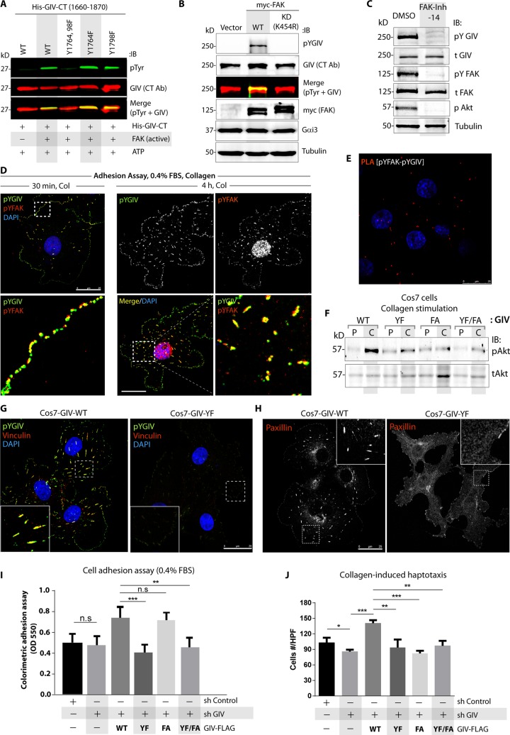
FIGURE 2:
Phosphorylation of GIV by FAK is required for cell–ECM adhesion and collagen-induced haptotaxis. (A) In vitro kinase assays were carried out with recombinant FAK and equal aliquots of either WT or mutant His-GIV-CT (aa 1660–1870) proteins. Phosphorylated GIV was detected by immunoblotting using anti–phospho-Tyr antibody (top; green). His-GIV-CT proteins were visualized using GIV-CT antibody (middle; red). Yellow pixels in the merged panel represent tyrosine-phosphorylated GIV-CT. (B) Cos7 cells expressing control vector, wild-type FAK (myc-FAK-WT), or a kinase-dead mutant FAK (myc-FAK-KD) were analyzed by immunoblotting for phosphorylation of endogenous GIV using anti–phospho-Tyr-1764-GIV (pYGIV) antibody. Expression of FAK (myc), Gαi3, and tubulin was analyzed by immunoblotting. (C) Cos7 cells were grown on culture dishes coated with poly-d-lysine, treated with FAK inhibitor 14 (FAK-Inh-14) or vehicle (dimethyl sulfoxide [DMSO]) for 24 h, and then seeded on collagen-coated dishes for 1 h before lysis and analyzed for total (t) and phosphoproteins (p) by immunoblotting (IB). (D) Cos7 cells were acutely stimulated with collagen before fixation (see Materials and Methods) and stained with phospho–Tyr-1764-GIV (pYGIV; green), phospho–Tyr-397-FAK (pYFAK; red), and DAPI (nuclei; blue) and analyzed by confocal microscopy. Bar, 25 μm. (E) Cos7 cells were analyzed for interaction between active pYGIV and active pYFAK by in situ PLA using rabbit anti-pYGIV and mouse anti-pYFAK antibodies. Red dots indicate sites of interaction. Incubation with secondary antibodies alone or with paxillin antibody showed no signal (negative controls; Supplemental Figure S2A). Bar, 25 μm. (F) GIV-depleted Cos7 cells stably expressing FLAG-tagged WT or mutant GIV constructs were grown on culture dishes coated with poly-d-Lysine (P) and then seeded on collagen-coated (C) dishes for 1 h before lysis and analyzed for phospho (p-) and total (t-) Akt by immunoblotting (IB). Compared to GIV-WT cells, percentage phosphorylation of Akt was suppressed by 65% in GIV-YF cells, 82% in GIV-FA cells, and 73% in GIV-YF/FA cells (p < 0.001), as determined by band densitometry using LiCOR Odyssey. (G, H) GIV-depleted (sh GIV) Cos7 cells stably expressing GIV-WT or GIV-YF mutant were fixed and stained for phospho–Tyr-1764-GIV (pYGIV; green), vinculin (red), and DAPI (nuclei; blue; G) or for paxillin (red, shown in grayscale; H) and analyzed by confocal microscopy. Bar, 25 μm. (I) Colorimetric adhesion assays were carried out using control (sh Control), GIV-depleted (sh GIV), or GIV-depleted cells stably expressing various GIV constructs in media with low serum. Error bars represent mean ± SD; n = 3; **p < 0.01; ***p < 0.001; n.s, not significant. See also Supplemental Figure S2, B and C, for immunoblots showing the levels of GIV expression and other FA proteins in these cell lines. (J) Cells in I were analyzed for collagen-induced haptotactic cell motility using Transwell assays. Images of representative fields are displayed in Supplemental Figure S2D. Bar graphs show quantification of the number of cells that migrated averaged from ∼ 20 high-power field-of-view images per experiment. Error bars represent mean ± SD; n = 3; *p < 0.05, **p < 0.01, ***p < 0.001.

