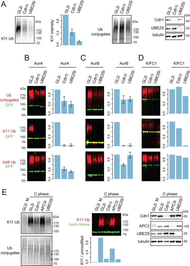
FIGURE 4:
Regulation of K11 chain assembly by APC/C-Cdh1. (A) U2OS-bioUb cells transfected with siRNA sequence against GL2 (control), Cdh1, or UBE2S were synchronized to mitotic exit. Whole-cell lysate was interrogated by K11 linkage–specific antibody and by biotin antibody to show total ubiquitin conjugates. (B–D) U2OS-bioUb cells were transfected with indicated Venus-tagged constructs together with control (GL2) or Cdh1 or UBE2S siRNA sequence, induced for bioUb expression for 44 h, and synchronized to mitotic exit. Cellular ubiquitination assays were carried out to detect total ubiquitin, K11 linkage, and K48 linkage on each substrate. Bar plot shows mean measurements from three repeats for AurA and AurB and two repeats for KIFC1, with SD plotted where three repeats are available. In vivo degradation assays were carried out in parallel (shown in Supplemental Figure S4), together with representative blots to validate depletions. (E) U2OS-AurA-Venus cells were transfected with indicated siRNAs and synchronized to prometaphase using a sequential thymidine and STLC block. Cells were then released into mitotic exit by treating with 300 nM CDK I/II inhibitor for 45 min before harvesting. Whole-cell lysates (left) or AurA-Venus pull downs (middle) were probed for ubiquitin conjugates. Whole-cell lysates were also tested for knockdown efficacies with indicated antibodies (right).

