FIGURE 1:
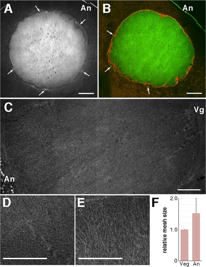
Structure of the intranuclear actin filaments. Eleven oocytes derived from four different females were examined. The nucleus of a midsagittal (animal–vegetal) cryosection of a full-grown stage VI oocyte was stained with Alexa 488–phalloidin (A) and double stained with Alexa 488–phalloidin and anti-lamin antibody (B). Arrows indicate the actin filaments surrounding the nucleus. (C) Enlarged image of the nucleus by assembling three shots, which had to be taken to cover one section, in a composite plate. The vegetal region (D) and animal region (E) are further enlarged. (F) Comparison of the actin filament mesh size between the vegetal (Veg) and animal (An) sides. The area of the space surrounded by actin filaments (the mesh hole) was measured over a set range by ImageJ software. Twelve oocytes from nine different females were measured. Relative mesh size at the animal side. Bars, 100 μm (A, B), 50 μm (C–E). An, animal pole; Vg, vegetal pole.

