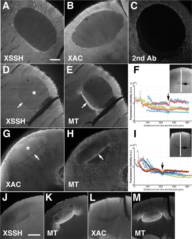
FIGURE 4:
Localization of XSSH and XAC during oocyte maturation. Midsagittal sections of full-grown oocytes (A, B) were stained with anti-XSSH antibody (A) and anti-XAC antibody (B). For the negative control, the cryosection of the oocyte at the relative time point of 0.8 was stained with Alexa 488–labeled secondary antibody against rabbit IgG alone (C). Midsagittal sections of maturing oocytes just after GVBD (D–I) and at the relative time point of 1.0 (J–M) were double stained with anti-XSSH antibody (D, J) and anti-tubulin antibody (E, K) or with anti-XAC antibody (G, L) and anti-tubulin antibody (H, M). At the relative time point of 1.0 (at the same time point as in J and L), the fluorescence intensity of either XSSH (F) or XAC (I) staining from the animal to the vegetal direction at the center of the nuclear region (shown with the white line in insets) was examined in different oocytes (six for XSSH and eight for XAC) using ImageJ software. The abscissa of the graphs is the number of pixels from the animal side, and the ordinate represents the fluorescence intensity (arbitrary units). Arrows indicate position of the base of the MTOC-TMA. The asterisks (D, G) indicate the nuclear region. Bars, 100 μm. Images are representative staining from at least 12 oocytes from four different females.

