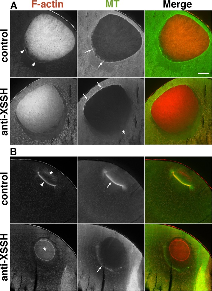
FIGURE 5:
Effects of injection of anti-XSSH antibody on assembly of the MTOC-TMA. Midsagittal sections of oocytes just before (A) or after (B) GVBD, injected with buffer alone (control) or with 10 mg/ml of 1:1 mixture of anti-XSSH IgG and anti-XSSH IgG-NLS (anti-XSSH), were double stained with TMR–phalloidin (F-actin) and anti-tubulin antibody (MT). (A) Assembly of the cytoplasmic actin filaments (arrowheads) and microtubule bundles (arrows) at the basal region of nuclei is clearly visible in the control but faint at the basal region of nuclei in antibody-injected oocytes (asterisk). On the other hand, microtubule bundles are evident at the animal side of the nuclei of antibody-injected oocytes. (B) The cytoplasmic actin filaments (arrowhead) are apparent at the base of the MTOC-TMA (arrow) in the control, whereas the intranuclear actin filaments (asterisks) clearly remained in a globular shape and the MTOC-TMA is faint at the basal region of the nuclei in antibody-injected oocytes. Merged images are also shown. Bar, 100 μm. Images in A and B are representative staining of 11 and 17 oocytes from five females, respectively.

