FIGURE 8:
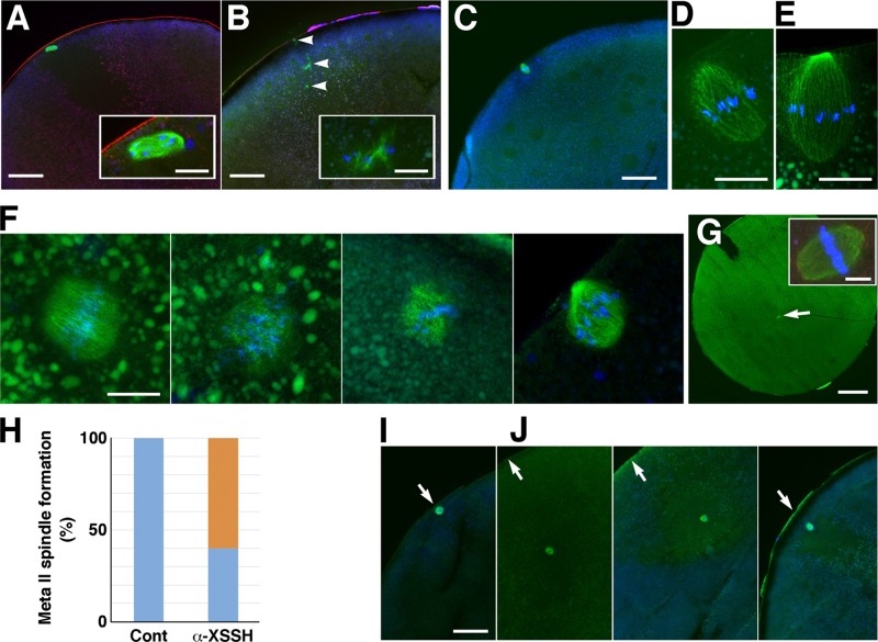
Effects of injection of anti-XSSH antibody (A–H) and S3A-cofilin (I, J) on assembly of meiotic spindles. Cryosections were triple stained with DAPI (blue), anti-microtubule antibody (green), and TMR-phalloidin (red; A, B, G inset) or double stained with DAPI (blue) and anti-microtubule antibody (green; C–G, I, J). (A) Control oocytes formed the metaphase I spindle (exactly prometaphase I) at the animal cortex (bar, 100 μm). The spindle is enlarged in the inset (bar, 20 μm). (B) Malformation of the metaphase I spindle is observed in the antibody-injected oocytes (bar, 100 μm). Arrowheads indicate disrupted microtubule bundles associated with chromosomes. Inset, enlarged image of one of the bundles (bar, 20 μm). (C–E) Representative images of metaphase II spindles oriented vertically to the cortex in the control oocytes. The spindle in C (bar, 100 μm) is enlarged in D (bar, 20 μm). Another example of metaphase II spindles is shown in E (bar, 20 μm). (F) Four examples of metaphase II spindles formed in the antibody-injected oocytes (bar, 20 μm). (G) An example of metaphase II spindles (arrow) formed at the center of the antibody-injected oocytes without anchoring to the cortex (bar, 200 μm). Inset, enlarged image of the spindle (bar, 20 μm). (H) Ratio of metaphase II spindles formed at the cortex (blue) to spindles formed at the center of oocytes (orange) in control (n = 9) and anti-XSSH antibody–injected oocytes (n = 10). (I, J) Metaphase I spindles formed at the cortex in a control oocyte (I) and formed in the yolk-free region without anchoring to the cortex in S3A-cofilin–injected oocytes (J; three examples are shown). Bar, 100 μm.

