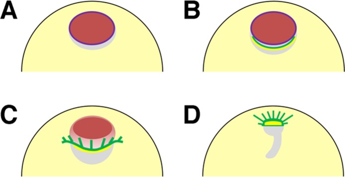
FIGURE 9:

Summary of organization of microtubules and actin filaments during oocyte maturation in Xenopus oocytes. (A) Stage VI oocytes form dense intranuclear actin filament networks (red) and cytoplasmic actin filament bundles surrounding the nuclei (not shown). Purple line represents nuclear envelopes (lamin). (B) As maturation begins, microtubules and cytoplasmic actin filaments (green and yellow, respectively) concentrate to the perinuclear cytoplasm (yolk-free zone, gray) at the vegetal side of the nuclei in a process that is dependent on XAC-mediated actin dynamics; this process is inhibited by anti-XSSH antibody injection. (C) When GVBD occurs from the vegetal side of the nuclei, the nucleus begins to shrink, the MTOC-TMA (green) develops well, and the cytoplasmic actin filaments (yellow) remain at its base. The intranuclear actin filaments then begin to disassemble from the vegetal side of the nuclei (orange). Although the TMA elongates to the animal side, it never enters the animal region, where the residual intranuclear actin filaments (red) are still present. (D) Disassembly of intranuclear actin filaments leads to the migration of the MTOC-TMA to the animal side and subsequent formation of the first meiotic spindles. The gray region represents the yolk-free zone.
