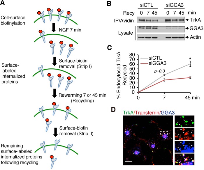
FIGURE 3:
GGA3 is required for TrkA recycling. (A) Schematic of internalization assay. PC12 (615) cells were biotinylated at 4°C and stimulated with NGF for 7 min at 37°C to allow for internalization. After glutathione stripping of the remaining cell-surface biotin, cells were reincubated at 37°C for another 7 or 45 min to allow for the recycling of internalized receptors. Cells were then stripped again with glutathione to remove biotin from the surface-exposed biotinylated receptors recycled back to the cell surface. The remaining biotinylated proteins were then collected with avidin and immunoblotted with TrkA antibodies. The TrkA signal lost in the second stripping procedure was considered the fraction of recycled receptors. (B) Representative Western blots of the TrkA recycling assay performed in control and GGA3-depleted PC12 (615) cells. Recy 0 refers to the internalized biotinylated receptors before rewarming; Recy 7 and 45 refer to the remaining biotinylated receptors after rewarming. (C) Quantification of the degree of TrkA recycling from three independent experiments (as described in A and B). The amount of recycled TrkA is expressed as the percentage of the pool of biotinylated TrkA following the 7-min internalization period and before rewarming. Student’s t test, *p < 0.05. (D) Confocal microscopy images comparing the distribution of endogenous GGA3 with that of cell surface–labeled TrkA (5C3 antibodies) and transferrin receptor (Alexa Fluor 594–conjugated transferrin) internalized for 15 min in PC12 (615) cells. Insets, regions of higher magnification; arrowheads indicate colocalization. Scale bar, 10 μm.

