FIGURE 6:
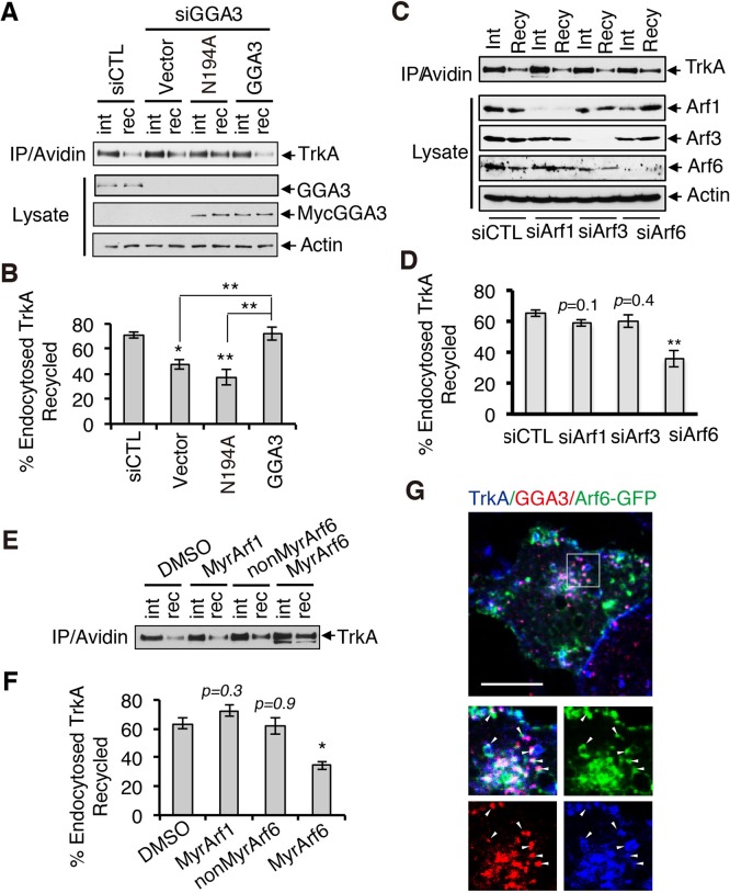
GGA3-mediated TrkA recycling requires Arf6. (A) Analysis of TrkA recycling in GGA3-depleted PC12 (615) cells rescued with control cDNA (vector) or siRNA-resistant GGA3 wild type or GGA3 mutant unable to bind Arf-GTP (GGA3-N194A). Recycling assays were performed as described in Figure 3A. int, internalized biotinylated TrkA before rewarming; rec, remaining biotinylated receptors after 45 min of rewarming (recycling). (B) Quantification of the degree of TrkA recycling from three independent experiments (as described in A). The amount of recycled TrkA is expressed as the percentage of the pool of biotinylated TrkA after the 7-min internalization period and before rewarming. One-way ANOVA, *p < 0.05, **p < 0.01. (C) Analysis of TrkA recycling in PC12 (615) cells treated with control, Arf1, Arf3, and Arf6 siRNA. Recycling assays were performed as described in Figure 3A. int, internalized biotinylated TrkA before rewarming; rec, remaining biotinylated receptors after 45 min of rewarming (recycling). (D) Quantification of the degree of TrkA recycling from three independent experiments (as described in C). The amount of recycled TrkA is expressed as the percentage of the pool of biotinylated TrkA after the 7-min internalization period and before rewarming. One-way ANOVA, **p < 0.01. (E) Analysis of TrkA recycling in PC12 (615) cells treated with dimethyl sulfoxide (vehicle), MyrArf1, MyrArf6, and NonMyrArf6 (nonpermeant Arf6 peptide control). Recycling assays were performed as described in Figure 3A. int, internalized biotinylated TrkA before rewarming; rec, remaining biotinylated receptors after 45 min of rewarming (recycling). (F) Quantification of the effects of MyrArf1, MyrArf6, and NonMyrArf6 peptides on the degree of TrkA recycling from three independent experiments (as described in A). One-way ANOVA, *p < 0.05. (G) Confocal microscopy images comparing the distribution of Arf6-GFP, endogenous GGA3, and cell surface–labeled TrkA receptors (5C3 antibodies) internalized for 15 min in PC12 (615) cells. Insets, regions of higher magnification; arrowheads indicate TrkA-labeled vesicles. Scale bar, 10 μm.

