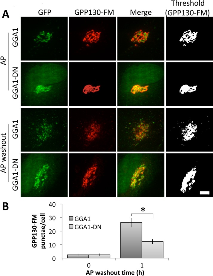
FIGURE 3:
GGA1-DN blocks GPP130-FM trafficking. (A) Cells were cotransfected with HA-tagged GPP130-FM and either GFP-tagged GGA1 or GGA1-DN and then either left in AP or treated with a 1 h of AP washout. Images show GFP fluorescence from the transfected proteins and anti-HA staining, with the final panel using uniform thresholding of the GPP130-FM image to aid visualization of small punctae. Bar, 5 μm. (B) Quantified appearance of GPP130 in peripheral punctae of cells expressing GGA1 or GGA1-DN after 0 or 1 h of AP washout (mean ± SEM, n = 3, >10 cells/experiment, *p < 0.05).

