FIGURE 5:
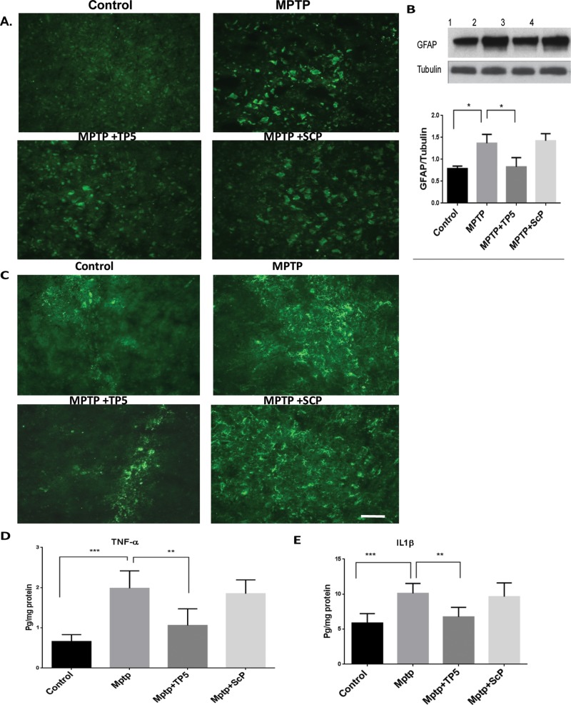
TP5 suppresses MPTP-induced astroglial and microglial activation and inflammation in the SN in vivo. (A) Sections of SN tissues obtained from the same animals as used in Figure 3 were immunostained with GFAP antibody for astrocyte and Cdllb (C) for microglia. The heightened expression of GFAP and CdIIb was observed in the MPTP group as compared with the control group, whereas the MPTP group treated with TP5 reveals a moderate staining of Cd11b and GFAP. The control group, however, shows almost negligible staining. Scale bar, 200 mm. (B) Western blots of tissue lysates from each group show a marked reduction in the expression of MPTP-induced GFAP by TP5. In addition, sandwich ELISAs of lysates from each group also show a TP5 rescue of overexpressed TNF-α (D) and IL-1β (E) in MPTP-induced inflammation (n = 8, ***p < 0.001, **p < 0.01).

