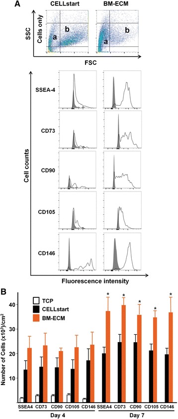
Fig. 4.

Phenotypic expression of MSC surface markers after culture in SFM for 4 and 7 days. Early passage (P1) cultures of BM-MSCs were grown on TCP, BM-ECM, or CELLstart™ in SFM. Phenotypic expression of MSC-associated markers (SSEA-4, CD73, CD90, CD105, and CD146) was assessed by using flow cytometry. a Single-cell suspensions, derived from 7-day cultures on CELLstart™ or BM-ECM, were analyzed by fluorescence-activated cell sorting. In the top panel, dot plots of the cell distribution are shown. Relatively smaller cells are found in “range a” (CELLstart: approximately 30 %; BM-ECM: 62 %), whereas relatively larger cells are found in “range b” (CELLstart: approximately 35 %; BM-ECM: 7 %). In the lower panel, histograms represent the expression of the indicated markers. Cells were stained with primary non-specific antibody (isotype, IgG) as negative controls (gray peaks). b P1 cultures of BM-MSCs were grown on the three culture surfaces for 4 (left panel) and 7 (right panel) days in SFM. The number of positive cells expressing each marker was determined as a percentage of the total cell population (also see Table 1). Mean ± standard deviation was calculated from three independent experiments. *P < 0.05 versus CELLstart™. BM-ECM bone marrow-derived extracellular matrix, BM-MSC bone marrow-derived mesenchymal stem cell, CD, cluster of differentiation/determinants, FSC forward scatter, MSC mesenchymal stem cell, P1 passage 1, SFM serum-free media, SSC side scatter, SSEA-4 stage-specific embryonic antigen-4, TCP tissue culture plastic
