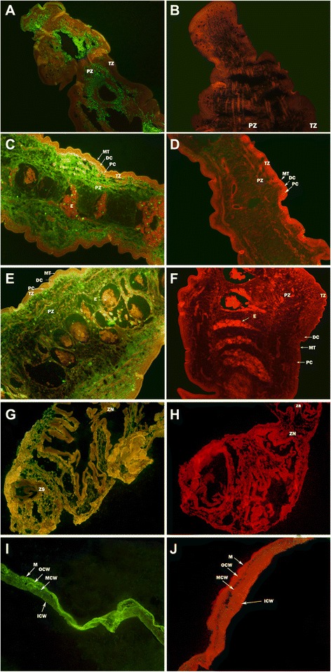
Fig. 2.

Immunolocalization of P2 protein in T. multiceps. The green fluorescence tint shows the location of TmP2 protein. a, antisera in cephalomeres of adult tapeworm; b, negative sera in cephalomeres of adult tapeworm; c, antisera in immature segments of adult tapeworm; d, negative sera in immature segments of adult tapeworm; e, antisera in mature segments of adult tapeworm; f, negative sera in mature segments of adult tapeworm. g, antisera in cephalomeres of coenurus; h, negative sera in cephalomeres of coenurus; i, antisera in cystic wall of coenurus; j, negative sera in cystic wall of coenurus. The magnification of all images is × 200. Arrows indicate the areas of the parasite: MT, microthrix; DC, distal cytoplasm; PC, perinuclear cytoplasm; TZ, tegument zone; PZ, parenchymatous zone; E, eggs; ZS, zone of scole; ZN, zone of neck; M, microtrichia; OCW, the outer layer of cystic wall; MCW, the middle layer of cystic wall; ICW, the inside layer of cystic wall
