
Fig. 2.
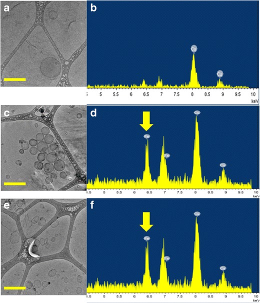
Cryo-TEM images (a) and EDS scans (b) of liposomes containing DOX, RAL, and P84 which show absence of iron peak at 6.4 keV. Cryo-TEM images and EDS scans of liposomes containing DOX, RAL, P84, and MNPs before (c, d) and after (e, f) RF exposure. The L/N ratio was 10,000:1 for these formulations. Yellow arrows indicate the presence of MNPs in the bilayers. The round shape of liposomes was converted to angular showing the influence of RF heating on the bilayers. Scale bar is 200 nm