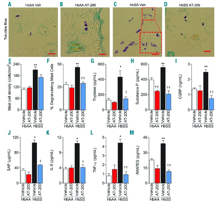
Figure 5.

AT-200 treatment reduces mast cell degranulation and inflammation. Control and sickle mice were treated with vehicle or 10 mg/kg AT-200/day for seven days as described in Figure 3D–E, and their dorsal skin and serum/plasma were analyzed 24 h following the last injection on day 7 (A–D). Toluidine blue staining for mast cells in dorsal skin: Inset in (C) shows magnified degranulating mast cells. Original magnification X900; scale bar, 20 mm. Each image represents 12–16 reproducible images per mouse from 10–12 mice per treatment. Image acquisition information: Olympus IX70 microscope, 60x/0.45 objective lens, DP70 digital camera and DP70 Manager Software (Olympus) and Adobe Photoshop. (E) Mast cell density shown as means ± SEM per square millimeter of skin sections stained with Toluidine blue. (F) Percentage of degranulating mast cells shown as means ± SEM degranulating mast cells to the total number of mast cells per square millimeter. Plasma concentrations are shown in (G) Tryptase, (H) Substance P, (I) CGRP, (J) SAP, (K) IL-6, (L) TNF-α, and (M) RANTES.. Statistical significance was calculated by comparing each value to control HbAA-BERK mice (*) and between vehicle and AT-200 (†). *P<0.05 and **P< 0.005 compared to vehicle-treated HbAA-BERK; and †P<0.05 and ††P<0.005 compared to vehicle for that time point. Mean age of mice ± SEM in months were, HbAA-BERK Vehicle (n=4), 4.06 ± 0.29, HbAA-BERK AT-200 (n=4), 4.04 ± 0.29, HbSS-BERK Vehicle (n=4–7), 4.38 ± 0.32, and HbSS-BERK AT-200 (n=4–7), 4.41 ± 0.34. Veh: vehicle; CGRP: Calcitonin Gene-Related Protein; SAP: Serum Amyloid Protein; IL-6: Interleukin 6; TNF-α: Tumor Necrosis Factor alpha; RANTES: Regulated on Activation, Normal T-Cell Expressed and Secreted.
