
Figure 7.
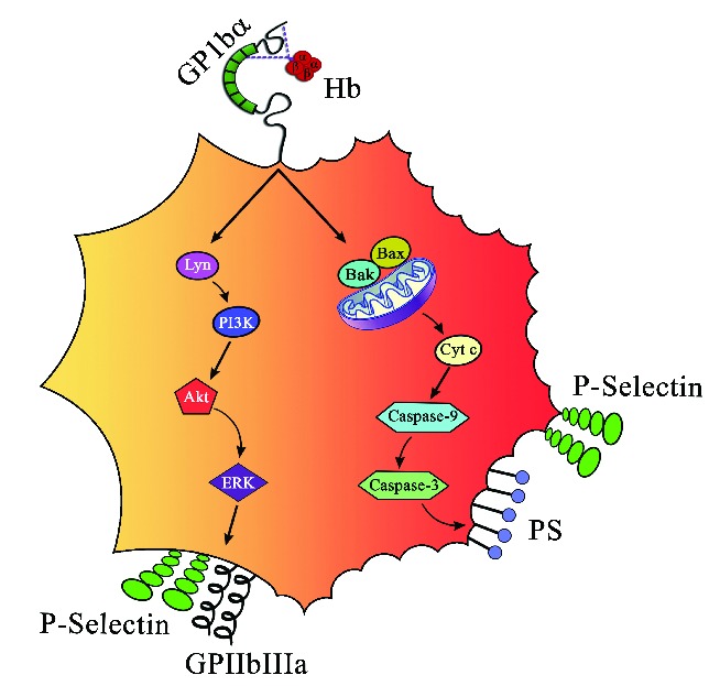
Schematic representation of platelet activation and apoptosis by Hb. Hb at lower concentrations (0.37–3 μM) induces inside out signaling via Lyn, PI3K, AKT, and ERK by binding to GP1bα and increases surface expression of P-selectin and GPIIb-IIIa. On the other hand, concentrations of Hb (3-6 μM) induce apoptosis and increase expression of Bak and Bax, release cytochrome C, activate caspase-9 and caspase-3, and expose PS on platelets.