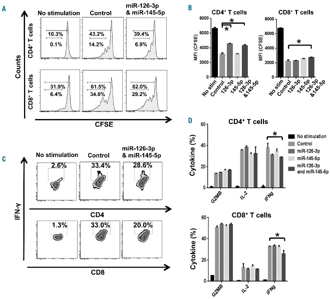
Figure 7.
Overexpression of miR-126-3p and miR-145-5p in T cells from AA patients decreases T-cell proliferation and inhibits IFN-γ production. Purified CD4+ and CD8+ T cells from AA patients were labeled with CFSE and transfected with miR-126-3p and/or miR-145-5p, followed by stimulation with anti-CD3/CD28 beads. (A) Representative histograms of CD4+ and CD8+ T-cell proliferation. (B) Frequency of CD4+ or CD8+ T-cell proliferation was quantified as the median fluorescence intensity (MFI) of CFSE from dividing cells, after transfection of control, miR-126-3p, miR-145-5p, or both miRNA. *P<0.05 [one-way analysis of variance (ANOVA)]. (C) Representative plots of intracellular IFN-γ expression following in vitro stimulation with anti-CD3/CD28 beads. (D) Percentages of T cells producing GZMB, IFN-γ, and IL-2 were examined in CD4+ or CD8+ T cells transfected with miR-126-3p and/or miR-145-5p 48 h later. Data are from three independent experiments (means ± SEM). *P<0.05. [two-way analysis of variance (ANOVA)].

