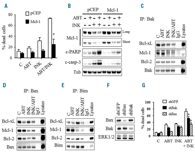
Figure 6.

Functional role of Mcl-1, Bax, and Bak in INK128/ABT-737-mediated cell death. A) U937 cells ectopically expressing Mcl-1 and their empty vector control pCEP cells were exposed to INK128 (200 nM) ± ABT-737 (500 nM) for 24 hrs, after which cell death was assessed using the Annexin V/PI staining assay. Error Bars: SD of 3 independent experiments; *P<0.01. Alternatively, protein lysates were prepared and subjected to Western blot analysis (B). (C–E) U937 cells were treated with INK128 (200 nM) ± ABT-737 (500 nM) for 4 hrs after which cells were lysed, Bak (C), Bax (D), or Bim (E) were immuno-precipitated, and the immuno-precipitates were subjected to immunoblotting. F) Western blot analysis in U937 cells in which Bax or Bak was knocked down using shRNA. G) These cells were exposed to INK128 (200 nM) ± ABT-737 (500 nM) for 24 hrs, after which cell death was monitored using the Annexin V/PI staining assay. Error Bars: SD of 3 independent experiments; *P<0.01 for shBax and P<0.05 for shBak.
