Abstract
Central nervous system involvement by malignant cells is a rare complication of Waldenström macroglobulinemia, and this clinicopathological entity is referred to as the Bing-Neel syndrome. There is currently no consensus on the diagnostic criteria, therapeutic approaches and response evaluation for this syndrome. In this series, we retrospectively analyzed 44 French patients with Bing-Neel syndrome. Bing-Neel syndrome was the first manifestation of Waldenström macroglobulinemia in 36% of patients. When Waldenström macroglobulinemia was diagnosed prior to Bing-Neel syndrome, the median time interval between this diagnosis and the onset of Bing-Neel syndrome was 8.9 years. This study highlights the possibility of the occurrence of Bing-Neel syndrome without any other evidence of progression of Waldenström macroglobulinemia. The clinical presentation was heterogeneous without any specific signs or symptoms. Biologically, the median lymphocyte count in the cerebrospinal fluid was 31/mm3. Magnetic resonance imaging revealed abnormalities in 78% of the cases. The overall response rate after first-line treatment was 70%, and the overall survival rate after the diagnosis of Bing-Neel syndrome was 71% at 5 years. Altogether, these results suggest that Bing-Neel syndrome should be considered in the context of any unexplained neurological symptoms associated with Waldenström macroglobulinemia. The diagnostic approach should be based on cerebrospinal fluid analysis and magnetic resonance imaging of the brain and spinal axis. It still remains difficult to establish treatment recommendations or prognostic factors in the absence of large-scale, prospective, observational studies.
Introduction
Waldenström macroglobulinemia (WM) is a rare, indolent B-cell lymphoproliferative disorder, classified as lymphoplasmacytic lymphoma in the 2008 World Health Organization classification. It has a wide spectrum of complications, mostly related to the monoclonal M-component (e.g. hyperviscosity syndrome, cryoglobulinemia, cold agglutinin hemolytic anemia, IgM-related neuropathies or tissue deposition). Neurological complications are dominated by IgM-related neuropathies (such as demyelinating peripheral neuropathy with IgM antibody activity against myelin-associated glycoprotein), but direct involvement of the central nervous system (CNS) by malignant lymphoid cells can occur. It was first described in 1936 by Jens Bing and Axel Neel, who reported two cases with hyperglobulinemia and CNS involvement,1 8 years before the first description of WM was reported by Jan Waldenström.2
Bing-Neel syndrome (BNS) is a rare and probably under-recognized complication of WM. Limited information is currently available in the literature, which is mostly based on case report descriptions. There is currently no consensus on the diagnostic criteria, treatment strategies and evaluation of response. BNS can present as either a diffuse or tumoral form. In the diffuse form, malignant cells infiltrate the leptomeningeal space, periventricular white matter or spinal cord. The tumoral form can be characterized by the presence of an intraparenchymal mass or nodular lesion. The distinction between these two forms is mainly based on imaging data.
We report the largest retrospective study to date in order to better characterize the clinical symptoms, biological features, radiological findings and clinical outcomes of patients with BNS.
Methods
Patients registered in the databases of 17 French centers were retrospectively analyzed in this multicenter, observational study. Patients were included if they had non-ambiguous cytological or histopathological evidence of CNS involvement by a lymphoplasmacytic proliferation, concomitant with a diagnosis of systemic WM according to the Second International Workshop on WM.3 We excluded all patients with a diagnosis of aggressive B-cell lymphoma resulting from the transformation of WM and patients presenting with neurological symptoms without clear cytological or histopathological evidence of CNS infiltration by lymphoplasmacytic cells. The study was approved by an independent ethics committee and was conducted in accordance with the Declaration of Helsinki.
The response criteria for BNS were defined as follows: complete remission when clinical symptoms disappeared with normalization of cerebrospinal fluid (CSF) and magnetic resonance imaging (MRI) findings; uncertain complete remission when clinical symptoms disappeared with either normalization of MRI but without CSF evaluation available at the end of the treatment, or normalization of CSF without MRI evaluation; and partial response when there was clinical, CSF or radiological partial improvement, including patients with neurological sequelae. Treatment failure was defined as no improvement or progression of clinical symptoms, CSF involvement or radiological findings.
Progression-free survival and overall survival were plotted using the Kaplan-Meier method, and the curves were compared using the log-rank test.
Results
Patients’ characteristics
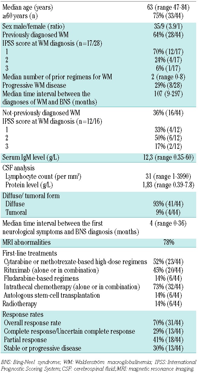
Forty-four patients treated for BNS between 1995 and 2014 were identified at 17 French centers. At the time of BNS diagnosis, the median age was 63 years (range, 47–84 years) and 35 patients (80%) were male. In 16 cases (36%), BNS was the first manifestation of WM; five of these patients had a previous diagnosis of monoclonal gammopathy of undetermined significance and the median time between the diagnosis of this gammopathy and that of BNS was 48 months (range, 9 to 108 months). For the 28 (64%) patients previously diagnosed with WM, the median time interval between the diagnosis of WM and that of BNS was 8.9 years (range, 9 months to 24.7 years). For 20 (71%) of the 28 patients previously diagnosed with WM, BNS occurred independently of a systemic progression of WM. Thirteen patients (33%, 13/39 patients with available data) were reported to have a diagnosis of peripheral neuropathy before the onset of BNS, and anti-glycolipid antibodies were positive in six (46%) of them [5 patients with anti-myelin-associated glycoprotein (anti-MAG) antibodies and 1 patient with anti-ganglioside (anti-GM1) antibodies]. The patients’ characteristics are summarized in Table 1.
Table 1.
Patients’ characteristics.
Clinical presentation of Bing-Neel syndrome
The median time interval between the appearance of neurological symptoms and the diagnosis of BNS was 4 months, with an upper limit of 36 months. The interval was longer than 1 year in nine (20%) patients. The symptoms or signs that led to the diagnosis of BNS were extremely heterogeneous, the most common being a balance disorder or disturbed gait [21 patients (48%), with ataxia described in 15 patients and dizziness in 6 patients] and cranial nerve involvement [13 patients (36%), with a predominance of facial or oculomotor nerve palsy]. Others ocular symptoms were decreased visual acuity or blurred vision. Other signs were poor performance status (>2) (12/44, 27%), cognitive impairment with frontal syndrome, memory loss or dementia (12/44, 27%), sensory deficit with hypoesthesia, dysesthesia or paresthesia (11/44, 25%), headache (8/44, 18%), pain (mainly localized in the back, neck or limbs) (8/44, 18%), cauda equina syndrome (6/44, 14%), motor deficit (6/44, 14%) and dysarthria or aphasia. Four patients with a tumoral form presented with convulsions, hemiparesis or aphasia. No intraocular involvement was documented in this series.
Biological results
CSF analysis was performed for all patients. The median lymphocyte count in CSF was 31 cells/mm3 (range, 1–3990). Monotypy, assessed by flow cytometry, could be confirmed in 31 (94%) of 33 cases with available data, with monotypic kappa light chain restriction in 84% and lambda restriction in 16% of the cases. The diagnosis of BNS for the 13 patients with no monotypy relied on the cytology of CSF or the histopathology of a CNS biopsy demonstrating the lymphoplasmacytic infiltration. The median protein level in CSF was 1.83 g/L (range, 0.39–7.8) and was increased (>0.4 g/L) in 39 (95%) cases. The diagnosis was assessed by the histopathology of a meningeal or a brain biopsy in eight cases. No data are available in this series regarding electrophoresis of CSF as this was not performed in routine practice.
At the time of the diagnosis of BNS, the median serum IgM level was 12.3 g/L (range, 0.35–60 g/L). Six patients had a concomitant immunophenotypic characterization of blood or bone marrow and CNS specimens, which was concordant in all cases.
Radiological findings
Magnetic resonance imaging was performed in 41 (93%) patients and was abnormal in 32 (78%) cases according to the local physicians’ interpretation. Seventeen patients had a cerebral computed tomography scan, which was abnormal in six (35%). All three patients who did not have MRI imaging had a normal cerebral computed tomography scan.
Two neuroradiologists reviewed the available MRI analysis of ten patients (all 10 patients had brain MRI analysis, and 7 of them had concomitant spine MRI analysis) before or immediately after the diagnosis of BNS. Brain parenchymal involvement was present in the classical sequences (characterized by high signal in T2 and iso- or hypointensity in T1 sequences) in six patients (6/10) with a predilection for sub-cortical or peri-ventricular locations. Medullary parenchymal involvement was found in two out of seven patients with medullary MRI imaging. One patient (1/10) had evidence of optic nerve involvement. A brain diffusion study was available for six patients; the diffusion sequence was normal in four patients and abnormal in two patients who showed cerebral vasogenic edema. Gadolinium injection also revealed cerebral or medullary leptomeningeal involvement in eight out of ten patients. Six out of seven patients with medullary MRI available had cauda equina enhancement after gadolinium injection. Finally, dura matter involvement, better visible after gadolinium injection, was present in six patients (6/10).
Treatment and response rates
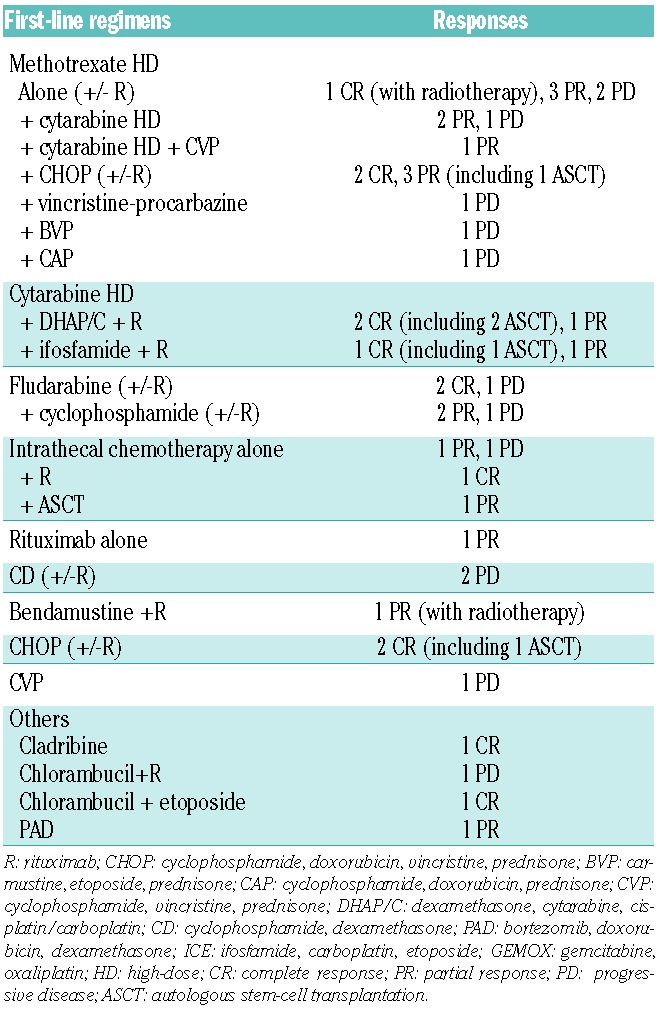
First-line treatment consisted of systemic chemotherapy in 40 (91%) cases and was based on high-dose chemotherapy in 52% of cases (methotrexate and/or cytarabine). Intrathecal chemotherapy was given to 32 (73%) patients. Fludarabine-based regimens were used in six patients (14%) (Table 2). Rituximab was part of the first-line treatment in 20 (45%) patients. Autologous stem-cell transplantation was performed as first-line therapy in six (14%) patients; conditioning regimens used were carmustine-etoposide-cytarabine-melphalan (BEAM) for two patients, bendamustine-etoposide-cytarabine-melphalan (Be-EAM), total body irradiation with melphalan, thiotepa-busulfan-cyclophosphamide and thiotepa-busulfan-melphalan. For six patients, the treatment was completed by whole-brain radiotherapy. Two patients received only intrathecal chemotherapy (only 1 patient responded). First-line treatments are detailed in Table 2 and responses according to first-line regimens are summarized in Table 3.
Table 2.
Chemotherapeutic regimens used.
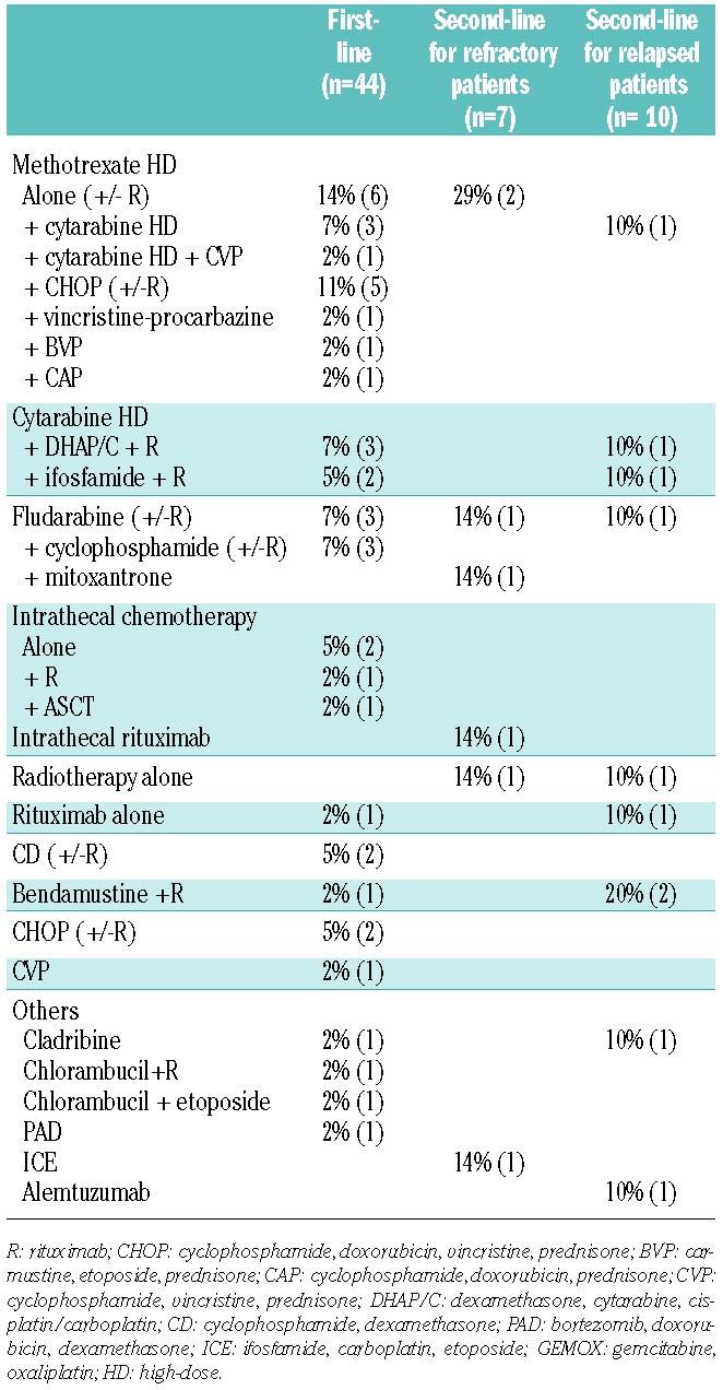
Table 3.
Responses according to first-line regimens.
After first-line treatment, the overall response rate of patients with BNS was 70% [complete response, n=1 (2%); uncertain complete response, n=12 (27%); partial response, n=18 (41%)]. Stable disease or progression was observed in 13 (30%) patients. All six patients who underwent autologous stem-cell transplantation responded, with four complete responses and two partial responses. After first-line treatment, the median serum M immunoglobulin level had decreased to 3 g/L. Among the 31 patients who responded to the first-line treatment, ten (30%) relapsed after a median of 16.5 months (range, 2–68 months), and seven (70%) responded to a second line of therapy. Seven of the 13 refractory patients underwent salvage therapy; only three of them (43%) responded and six patients died before salvage therapy could be initiated. Salvage treatments are summarized in Table 2.
We could not identify any difference in the response rates according to the first-line chemotherapy regimens used. The responses were heterogeneous but no predictive marker for treatment response based on biological parameters or chemotherapeutic regimens used could be identified.
Survival
The median follow-up period of living patients was 4.6 years after the diagnosis of BNS and 11.4 years after the diagnosis of WM. The overall survival rate after BNS diagnosis was 71% at 5 years and 59% at 10 years (Figure 1). The median overall survival from the time of the diagnosis of WM was 17.1 years. The median progression-free survival after the first-line treatment of BNS was 26 months (Figure 2). Fourteen (32%) patients died, nine due to BNS, one of BNS-treatment-related causes, one due to WM progression and three due to other causes (myocardial infarction, infection secondary to chronic obstructive pulmonary disease and death of unknown cause for one patient).
Figure 1.
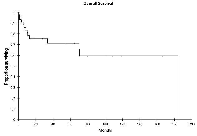
Overall survival of patients with BNS since their diagnosis.
Figure 2.
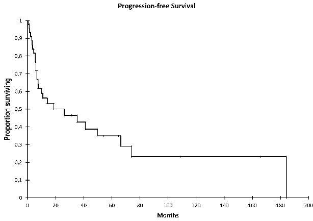
Progression-free survival of patients with BNS since their first-line treatment.
Discussion
To our knowledge, this series of patients with BNS is the largest ever studied. We used stringent inclusion criteria for all patients who had to have well-documented CNS involvement as confirmed by cytological and immunophenotyping analysis of CSF, or histopathological analysis of a brain biopsy. BNS is generally considered to be a rare complication of WM, but some cases are probably under diagnosed. This could be explained by the lack of specificity of clinical symptoms. A diagnosis of BNS should be considered in patients with WM in the case of any unexplained and persistent neurological manifestations. The first symptoms appeared in this series at a median of 4 months before the BNS diagnosis, although the delay was longer than 1 year for 20% of the patients. Another critical point that could explain the under recognition of BNS is the frequent occurrence of BNS independently of any systemic progression of WM (70% of cases previously diagnosed with WM in this series). In one third of the cases, BNS was the first manifestation of WM, and this study also highlights the possibility of a very late occurrence of BNS (up to 25 years after the diagnosis of WM). In our series, the incidence of peripheral neuropathies among the patients was 33% (as expected in a global population of WM patients). This represents another major pitfall in the diagnosis of BNS as peripheral neuropathy can mimic the symptoms of BNS (ataxia, sensory or motor deficit, pain), and an initial attribution of symptoms to peripheral neuropathy could delay the diagnosis of BNS. Thus BNS should be considered even in patients with a previously diagnosed peripheral neuropathy who complain of worsening symptoms or who are not responsive to treatment. A differential diagnosis to consider is another rare syndrome: the CANOMAD syndrome, characterized by the association of a chronic ataxic neuropathy, ophthalmoplegia, the presence of IgM monoclonal protein and anti-diasialosyl antibodies.
The diagnostic approach to BNS should be based on the findings of a lumbar puncture and MRI imaging of the brain and spinal cord. The CSF must be analyzed promptly after its collection. The cytological examination should be associated with immunophenotyping by flow cytometry in order to confirm the monotypy and the B-cell origin of the malignant cells and to demonstrate an immunophenotypic profile compatible with a WM proliferation. Electrophoresis performed on the CSF could show the M-component, but this examination is not always performed in routine practice; it is not sufficient alone and not specific enough to formally assess the CNS infiltration by tumor cells. The imaging protocol for the brain study of a suspected or known BNS should include T1-based images before and after gadolinium enhancement, T2 and T2* sequences, diffusion sequences, as well as delayed FLAIR images after gadolinium enhancement. The assessment should be completed with the study of the medulla, covering the whole spine, including T1 images before and after gadolinium enhancement, as well as T2 images. These analyses should be repeated in the case of suspected BNS if the initial results are negative, as illustrated by two patients in this series for whom the diagnosis of BNS could only be made after a second CSF analysis, and following previous examples reported in the literature.4–6 The diagnosis of BNS in the absence of radiological abnormalities should be made with caution and assessed after a multidisciplinary discussion before starting the treatment. The evaluation of the response at the end of treatment needs to carefully re-evaluate brain and spinal MRI, as well as CSF clearance.
The treatments observed in this retrospective study were based on local physicians’ choice and their heterogeneity precludes definite conclusions regarding the best treatment strategy to use. Most of the patients received systemic chemotherapy, notably when BNS was associated with WM progression. We did not observe any impact on overall and progression-free survival related to the use of high-dose chemotherapy (including cytarabine or methotrexate). Only a few patients were treated with fludarabine-based regimens in this series. However, several previous reports have suggested that purine analogs are effective treatment for BNS,7 and recent work confirmed the usefulness of fludarabine in the therapeutic armamentarium.8 Despite lack of evidence that rituximab penetrates the CNS, we observed that this drug was used in nearly half of the cases but without any impact on the survival or response rate. Intrathecal chemotherapy was frequently administered with systemic chemotherapy and was the only specific treatment in two patients. Autologous stem-cell transplantation was performed in six cases as first-line treatment. All patients responded to transplantation, are without relapse and are still alive. One patient underwent autologous stem-cell transplantation as second-line therapy and initially responded, but this patient died due to toxicity of the transplant (septic shock during aplasia). Autologous stem-cell transplantation has also been previously reported in the literature,9–13 but toxic deaths are described so that transplantation should be considered only for suitable patients.
The overall survival after the diagnosis of BNS in this series compares favorably with previously published data.14 The onset of BNS did not appear to be associated with a more aggressive clinical course of WM in this series (70% of patients had an International Prognostic Staging System score of 1 at the time of WM diagnosis with a median overall survival from the time of WM diagnosis of 17.1 years).
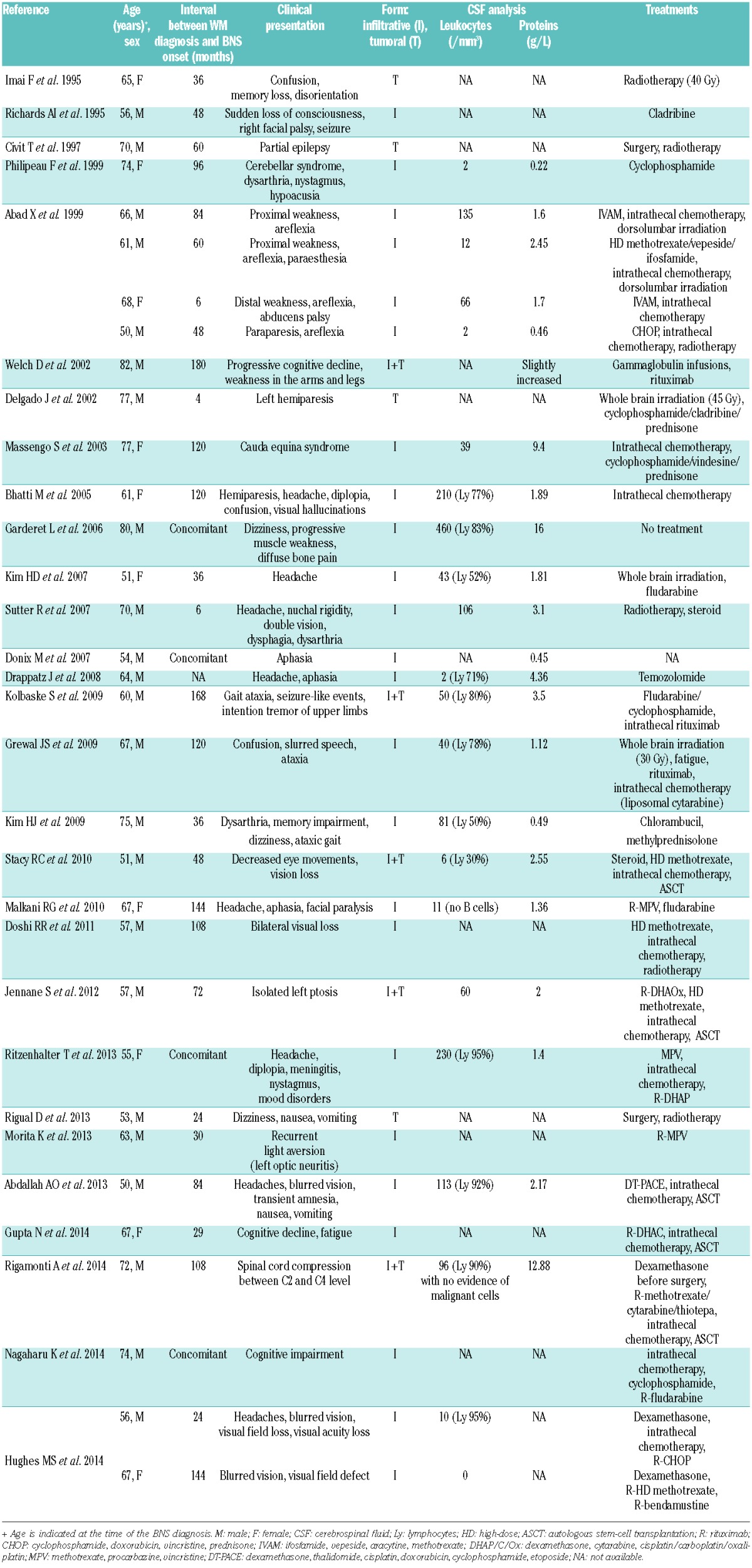
We found 33 cases published in the literature during the same period (1995–2014),4–7,9–13,15–35 which are some of the 56 cases described since the first description of BNS in 1936 (Table 4). The patients’ characteristics were similar: the median age at the time of BNS diagnosis was 65.5 years (range, 50–84), and the majority of patients were male (71%). Diffuse forms are predominant (74%), and the association of diffuse and tumoral forms was described in five cases. The median white blood cell count in CSF was 46.5/mm3 (range, 2–460) and the median protein level was 1.85 g/L (range, 0.22–16). As observed in our series, this review of the literature illustrates the possibility of a late onset of BNS, up to 25 years after the diagnosis of WM.
Table 4.
Review of the 33 cases of BNS published between 1995 and 2014.
The pathophysiology of BNS remains unexplained, but the role of hyperviscosity has been raised to explain the disruption of the blood-brain barrier. However, in our cohort, the median serum IgM level was rather low, and no patient experienced hyperviscosity before or at the time of BNS occurrence.
In summary, BNS is a rare and probably under recognized condition that should be considered early in the context of unexplained neurological signs in patients with WM. It can occur in patients with indolent and stable WM, leading to delays and potential pitfalls in establishing a prompt diagnosis. The treatment remains challenging, but it could be interesting to investigate the potential efficacy of ibrutinib in BNS patients as this kinase inhibitor has recently been demonstrated to produce a high response rate in relapsed or refractory WM.36 It was recently suggested that ibrutinib can penetrate the CNS on the basis of a recent study about mantle-cell lymphoma with CNS involvement.37 The diagnostic accuracy of BNS could be improved by the detection of the L265P mutation in the MYD88 gene in CSF specimens, as recently published.38 This test could be an interesting tool that will need further investigation to assess its potential utility for the diagnosis and evaluation of response to treatment in patients with BNS.
Acknowledgments
The authors would like to thank all the patients who participated in the study and the French association of WM patients (SILLC) for their continuing efforts to improve disease management and clinical research. The authors would also like to thank all the physicians from the Groupe d’Etude des Lymphomes Oculo-Cérébraux (LOC) for sharing data.
Footnotes
Authorship and Disclosures
Information on authorship, contributions, and financial & other disclosures was provided by the authors and is available with the online version of this article at www.haematologica.org.
References
- 1.Bing J, Neel AV. Two cases of hyperglobulinaemia with affection of the central nervous system on a toxi-infectious basis. Acta Med Scand. 1936;88(5–6):492–506. [Google Scholar]
- 2.Waldenström J. Incipient myelomatosis or “essential” hyperglobulinemia with fibrinogenopenia — a new syndrome? Acta Med Scand. 1944;117(3–4):216–247. [Google Scholar]
- 3.Owen RG, Treon SP, Al-Katib A, et al. Clinicopathological definition of Waldenstrom’s macroglobulinemia: consensus panel recommendations from the Second International Workshop on Waldenstrom’s Macroglobulinemia. Semin Oncol. 2003;30(2):110–115. [DOI] [PubMed] [Google Scholar]
- 4.Bhatti MT, Yuan C, Winter W, McSwain AS, Okun MS. Bilateral sixth nerve paresis in the Bing-Neel syndrome. Neurology. 2005;64(3): 576–577. [DOI] [PubMed] [Google Scholar]
- 5.Kolbaske S, Grossmann A, Benecke R, Wittstock M. Progressive gait ataxia and intention tremor in a case of Bing-Neel syndrome. J Neurol. 2009;256(8):1366–1368. [DOI] [PubMed] [Google Scholar]
- 6.Hughes MS, Atkins EJ, Cestari DM, Stacy RC, Hochberg F. Isolated optic nerve, chiasm, and tract involvement in Bing-Neel syndrome. J Neuroophthalmol. 2014;34(4): 340–345. [DOI] [PubMed] [Google Scholar]
- 7.Richards AI. Response of meningeal Waldenström’s macroglobulinemia to 2-chlorodeoxyadenosine. J Clin Oncol. 1995; 13(9):2476. [DOI] [PubMed] [Google Scholar]
- 8.Vos JM, Kersten M-J, Kraan W, et al. Effective treatment of Bing-Neel syndrome with oral fludarabine: a case series of four consecutive patients. Br J Haematol 2015. May 5 [Epub ahead of print] [DOI] [PubMed] [Google Scholar]
- 9.Stacy RC, Jakobiec FA, Hochberg FH, Hochberg EP, Cestari DM. Orbital involvement in Bing-Neel syndrome. J Neuroophthalmol. 2010;30(3):255–259. [DOI] [PubMed] [Google Scholar]
- 10.Jennane S, Doghmi K, Mahtat EM, Messaoudi N, Varet B, Mikdame M. Bing and Neel syndrome. Case Rep Hematol. 2012;2012:845091. [DOI] [PMC free article] [PubMed] [Google Scholar]
- 11.Abdallah A-O, Atrash S, Muzaffar J, et al. Successful treatment of Bing-Neel syndrome using intrathecal chemotherapy and systemic combination chemotherapy followed by BEAM auto-transplant: a case report and review of literature. Clin Lymphoma Myeloma Leuk. 2013;13(4):502–506. [DOI] [PubMed] [Google Scholar]
- 12.Gupta N, Gupta S, Al Ustwani O, Pokuri V, Hatoum H, Bhat S. Bing-Neel syndrome in a patient with Waldenstrom’s macroglobulinemia: a challenging diagnosis in the face of normal brain imaging. CNS Neurosci Ther. 2014;20(10):945–946. [DOI] [PMC free article] [PubMed] [Google Scholar]
- 13.Rigamonti A, Lauria G, Melzi P, et al. A case of Bing-Neel syndrome presenting as spinal cord compression. J Neurol Sci. 2014;346(1–2):345–347. [DOI] [PubMed] [Google Scholar]
- 14.Drouet T, Behin A, Psimaras D, Choquet S, Guillevin R, Hoang Xuan K. [Bing-Neel syndrome revealing Waldenström’s macroglobulinemia]. Rev Neurol (Paris). 2010;166(1): 66–75. [DOI] [PubMed] [Google Scholar]
- 15.Imai F, Fujisawa K, Kiya N, et al. Intracerebral infiltration by monoclonal plasmacytoid cells in Waldenstrom’s macroglobulinemia–case report. Neurol Med Chir (Tokyo). 1995;35(8):575–579. [DOI] [PubMed] [Google Scholar]
- 16.Civit T, Coulbois S, Baylac F, Taillandier L, Auque J. [Waldenström’s macroglobulinemia and cerebral lymphoplasmocytic proliferation: Bing and Neel syndrome. Apropos of a new case]. Neurochirurgie. 1997;43(4):245–249. [PubMed] [Google Scholar]
- 17.Philippeau F, Honnorat J, Nighoghossian N, et al. [JC virus infection and lymphoplasmocytic infiltration of the central nervous system revealed by a cerebellar syndrome]. Rev Neurol (Paris). 1999;155(11):961–965. [PubMed] [Google Scholar]
- 18.Abad S, Zagdanski A-M, Brechignac S, Thioliere B, Brouet J-C, Mariette X. Neurolymphomatosis in Waldenström’s macroglobulinaemia. Br J Haematol. 1999;106(1):100–103. [DOI] [PubMed] [Google Scholar]
- 19.Welch D, Whetsell WO, Weil RJ. Pathologic quiz case. A man with long-standing monoclonal gammopathy and new onset of confusion. Central nervous system involvement by Waldenström macroglobulinemia-Bing-Neel syndrome. Arch Pathol Lab Med. 2002;126(10):1243–1244. [DOI] [PubMed] [Google Scholar]
- 20.Delgado J, Canales MA, Garcia B, Alvarez-Ferreira J, Garcia-Grande A, Hernandez-Navarro F. Radiation therapy and combination of cladribine, cyclophosphamide, and prednisone as treatment of Bing-Neel syndrome: case report and review of the literature. Am J Hematol. 2002;69(2):127–131. [DOI] [PubMed] [Google Scholar]
- 21.Massengo S, Riffaud L, Morandi X, Bernard M, Verin M. Nervous system lymphoid infiltration in Waldenström’s macroglobulinemia. A case report. J Neurooncol. 2003;62(3):353–358. [DOI] [PubMed] [Google Scholar]
- 22.Garderet L, Baudel JL, Cervera P, et al. “Indolent” Waldenstrom’s macroglobulinemia and a cerebrospinal fluid protein level of 16 g/L. Eur J Haematol. 2006;77(1):80–82. [DOI] [PubMed] [Google Scholar]
- 23.Kim HD, Shin KC, Cho HS, Kim MK, Lee KH, Hyun MS. Therapeutic experience of Bing-Neel syndrome associated with Waldenstrom’s macroglobulinemia. J Korean Med Sci. 2007;22(6):1079–1081. [DOI] [PMC free article] [PubMed] [Google Scholar]
- 24.Sutter R, Arber C, Tichelli A, Steck AJ, Czaplinski A. Cranial and peripheral neuropathy due to leptomeningeal infiltration in a patient with Waldenstrom’s macroglobulinemia. J Neurol. 2007;254(8):1122–1123. [DOI] [PubMed] [Google Scholar]
- 25.Donix M, Beuthien-Baumann B, von Kummer R, Gahn G, Thomas F, Holthoff V. Nonfluent aphasia in a patient with Waldenstrom’s macroglobulinemia. J Clin Neurosci. 2007;14(6):601–603. [DOI] [PubMed] [Google Scholar]
- 26.Drappatz J, Akar S, Fisher DC, Samuels MA, Kesari S. Imaging of Bing-Neel syndrome. Neurology. 2008;70(16):1364. [DOI] [PubMed] [Google Scholar]
- 27.Grewal JS, Brar PK, Sahijdak WM, Tworek JA, Chottiner EG. Bing-Neel syndrome: a case report and systematic review of clinical manifestations, diagnosis, and treatment options. Clin Lymphoma Myeloma. 2009;9(6):462–466. [DOI] [PubMed] [Google Scholar]
- 28.Kim H-J, Suh S-I, Kim JH, Kim B-J. Brain magnetic resolution imaging to diagnose bing-neel syndrome. J Korean Neurosurg Soc. 2009;46(6):588–591. [DOI] [PMC free article] [PubMed] [Google Scholar]
- 29.Malkani RG, Tallman M, Gottardi-Littell N, et al. Bing-Neel syndrome: an illustrative case and a comprehensive review of the published literature. J Neurooncol. 2010;96(3):301–312. [DOI] [PubMed] [Google Scholar]
- 30.Doshi RR, Silkiss RZ, Imes RK. Orbital involvement in Bing-Neel syndrome. J Neuroophthalmol. 2011;31(1):94–95. [DOI] [PubMed] [Google Scholar]
- 31.Ritzenthaler T, Leray V, Bourdin G, et al. Ventriculitis revealing Bing-Neel syndrome in a patient without Waldenstrom’s macroglobulinemia. Clin Neurol Neurosurg. 2013;115(1):82–84. [DOI] [PubMed] [Google Scholar]
- 32.Rigual D, Qiu J, Fenstermaker RA, Fabiano AJ. Tumoral Bing-Neel syndrome presenting as a cerebellar mass. Clin Neurol Neurosurg. 2013;115(6):823–826. [DOI] [PMC free article] [PubMed] [Google Scholar]
- 33.Morita K, Yoshimi A, Masuda A, Ichikawa M, Yatomi Y, Kurokawa M. Unique association of Waldenström macroglobulinemia with optic neuritis and monoclonal T cell expansion. Int J Hematol. 2013;98(2):247–249. [DOI] [PubMed] [Google Scholar]
- 34.Nagaharu K, Miyazami K, Imai H, et al. Successful treatment of Bing-Neel syndrome using combination therapy with fludarabine and rituximab. Rinsh Ketsueki. 2014;55(12):2423–2428. [DOI] [PubMed] [Google Scholar]
- 35.Lancellotti G, Cohen-Bittan J, Makdessi S, et al. Late-onset Bing-Neel syndrome associated with delirium and Lewy body dementia. J Am Geriatr Soc. 2014;62(11):2225–2227. [DOI] [PubMed] [Google Scholar]
- 36.Treon SP, Tripsas CK, Meid K, et al. Ibrutinib in previously treated Waldenström’s macroglobulinemia. N Engl J Med. 2015;372(15):1430–1440. [DOI] [PubMed] [Google Scholar]
- 37.Bernard S, Goldwirt L, Amorim S, et al. Promising results of ibrutinib in three patients with secondary central nervous system mantle cell lymphoma. Blood. 2014;124(21):3105–3105. [Google Scholar]
- 38.Poulain S, Boyle EM, Roumier C, et al. MYD88 L265P mutation contributes to the diagnosis of Bing Neel syndrome. Br J Haematol. 2014;167(4):506–513. [DOI] [PubMed] [Google Scholar]


